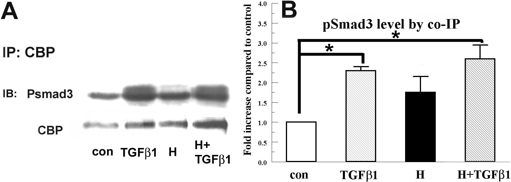
Figure 3. Hypoxia does not reduce TGFβ1
induced interaction between pSmads and CBP in rabbit keratocytes.
Nuclear extracts were collected 4 h after corresponding treatment and
immunoprecipitated with anti-CBP antibody. Eluates were separated by
SDS–PAGE and probed for pSmad3. Blots were also probed for CBP as an
internal control. A: Image shows a representative western blot.
B: Bar graph shows the relative change of pSmad3 over the
control group. Error bars represent the standard error of the mean
(n=3). The asterisk denotes that the indicated groups were
significantly different from control (p<0.05).

 Figure 3 of Xing, Mol Vis 2009; 15:1827-1834.
  Figure 3 of Xing, Mol Vis 2009; 15:1827-1834.