Figure 6. Cellular localization of the
p.L54P, p.N48K and WT CLRN1-HA polypeptides. The transfected BHK-21
cells were immunostained with HA antibody (red) showing the
localization of WT CLRN1-HA (E), the novel sequence alteration
p.L54P mutated CLRN1-HA (B) and the known disease-causing p.N48K
mutated CLRN1-HA (H). The same cells were immunostained with
plasma membrane –specific antibody (green) in panels A, D,
and G. Double-staining shows that WT CLRN1-HA (F) and
the p.L54P mutated CLRN1-HA (C) colocalize (yellow) with the
plasma membrane marker whereas the known mutation p.N48K (I)
does not colocalize with the plasma membrane marker. Cells were viewed
with a confocal immunofluorescence microscope. Scale bar represents 10
µm.
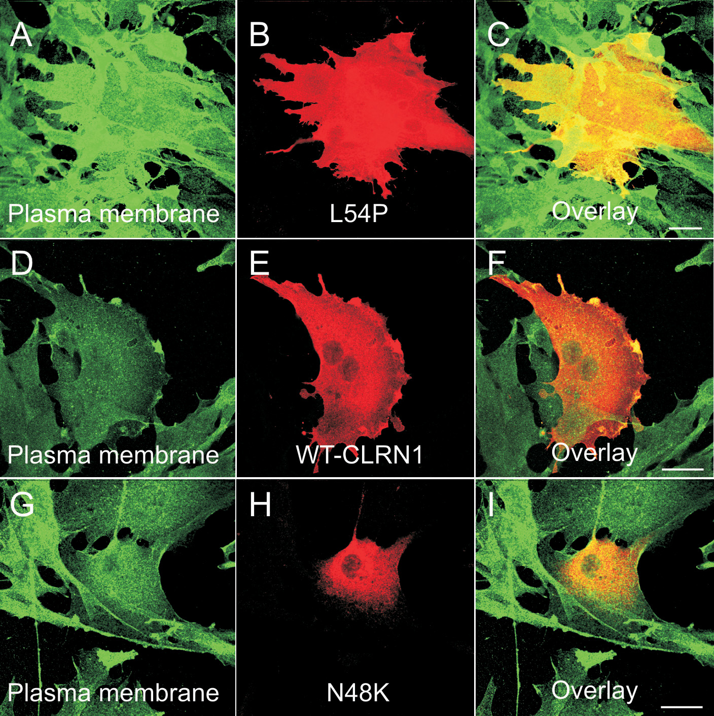
 Figure 6 of Isosomppi, Mol Vis 2009; 15:1806-1818.