Figure 5. Cellular localization of the
disease-causing mutant CLRN1-HA polypeptides. The transfected BHK-21
cells were double immunostained with HA antibody (red) in panels B,
E, H, K, and N showing the localization
of mutant CLRN1-HA. In panels A, D, G, J,
and M the cells were stained with the ER marker (green). The
right-most panels C, F, I, L, and O
show the overlay of the mutant CLRN1- HA staining (red) and ER-specific
staining (green). Yellow-orange staining indicates colocalization of
these stainings. Cells were viewed with a confocal immunofluorescence
microscope. Scale bar represents 10 µm.
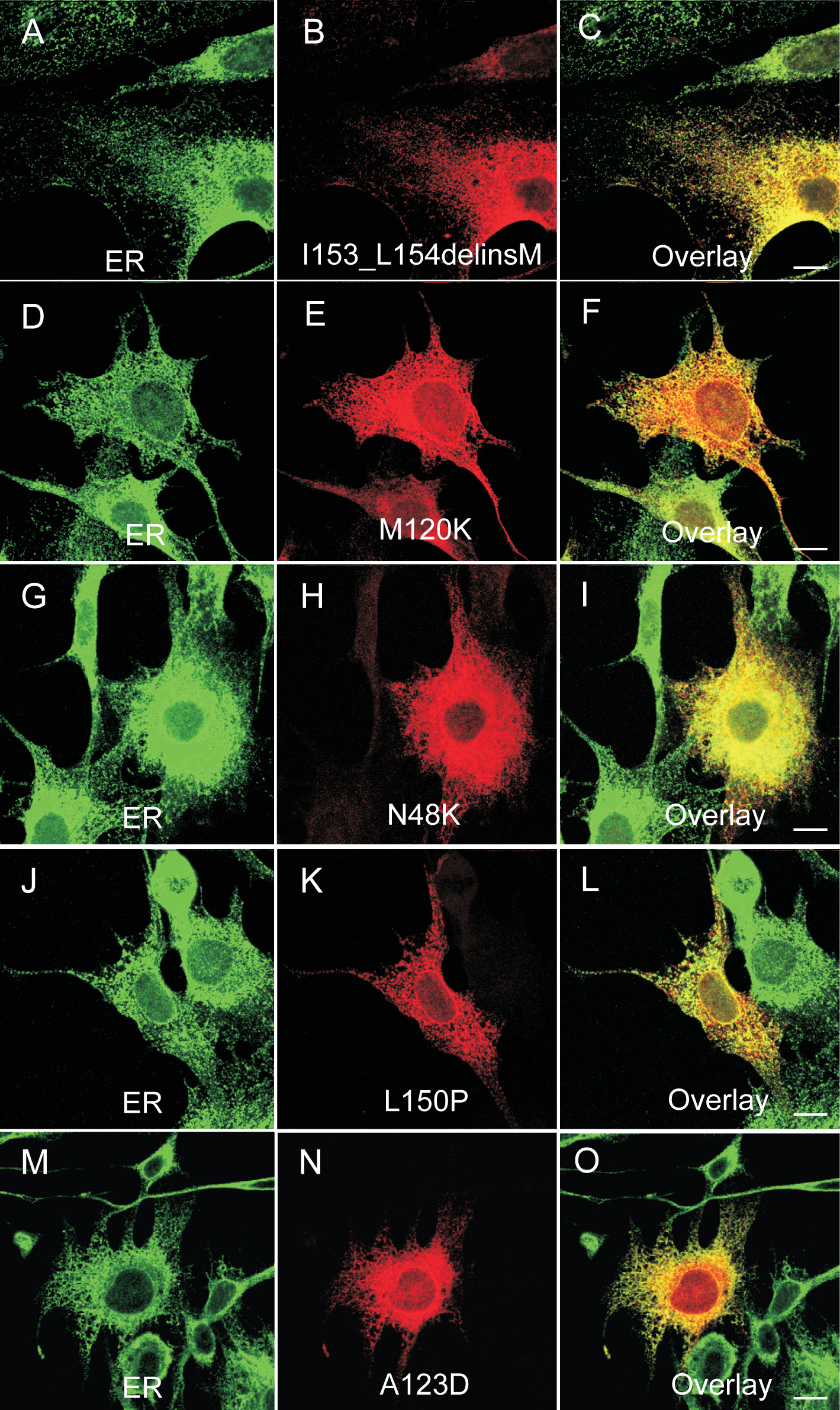
 Figure 5 of Isosomppi, Mol Vis 2009; 15:1806-1818.