Figure 4. Cellular localization of WT
CLRN1-HA protein in transfected BHK-21 cells. In panels B and E
the cells were immunostained with HA antibody (red). In panel A
the cells were immunostained with a plasma membrane specific antibody
(green) and in panel D with ER specific antibody (green). The
right-most panels (C and F) show the overlay of both
CLRN1-HA and the organelle-specific double staining. Yellow-orange
staining indicates an overlap of the CLRN1-HA protein (red) and
subcellular markers (green). Cells were viewed with a confocal
immunofluorescence microscope, magnification 63×. Scale bar represents
10 μm.
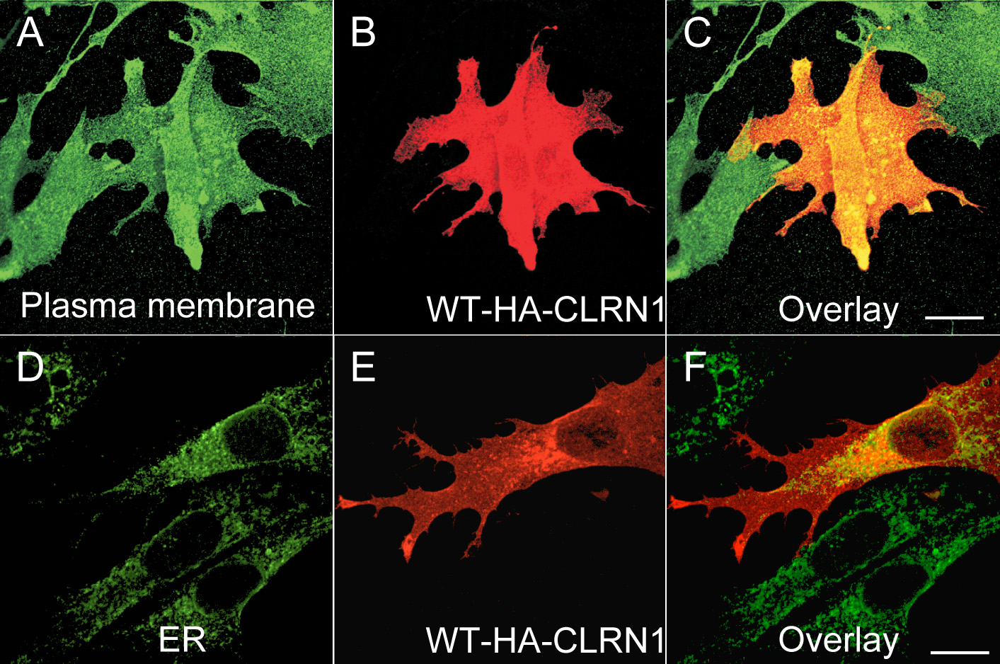
 Figure 4 of Isosomppi, Mol Vis 2009; 15:1806-1818.