Figure 1. Eye phenotype in Retinitis pigmentosa-nanophthalmos complex. A: Fundus photograph of the right eye from patient #1 reveals optic disc drusen, diffuse retinal pigment epithelium atrophy,
and blunting of the macular reflex. B: Fluorescein angiography shows choroidal transmission hyperfluorescence corresponding to retinal pigment epithelium atrophy
(patient #1, right eye). C: OCT image demonstrates cystoid macular edema, inner retinal layers splitting with discrete bridging elements at the fovea,
and macular cysts (patient #2, left eye).
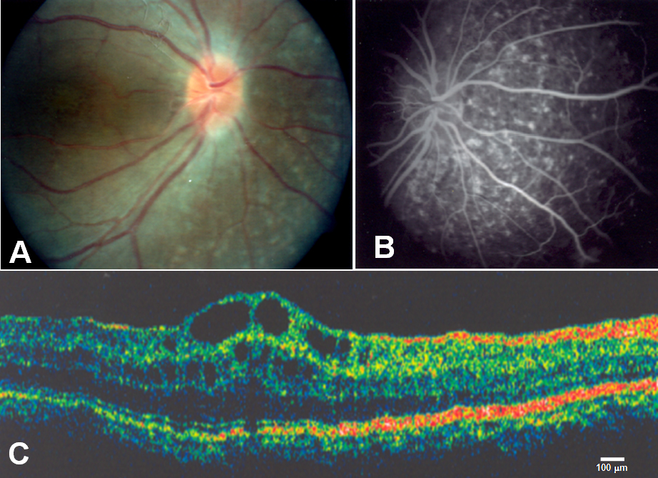
 Figure 1 of
Zenteno, Mol Vis 2009; 15:1794-1798.
Figure 1 of
Zenteno, Mol Vis 2009; 15:1794-1798.