Figure 3. Two ways to eliminate the Meta
III artifact. In A, dark and postbleach spectra were recorded
at acidic pH. The amount of Meta III formed was greatly reduced, and
its spectrum narrowed. Thus tracing rhodopsin diffusion at λ > 550
was only marginally compromised by Meta III formation. Spectra
represent average of six cells. In B, recordings were made in
standard Ringer at pH 7.5 with addition of 50 mM of freshly neutralized
hydroxylamine. Conversion of metaproducts to retinaloxime was complete
at 30 s postbleach, and afterwards metaproducts did not contribute to
absorbance changes. Spectra represent average of five cells.
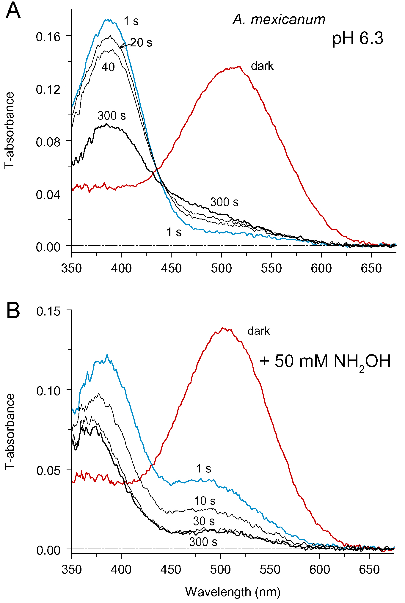
 Figure 3 of Govardovskii, Mol Vis 2009; 15:1717-1729.