Figure 2. Clinical finding’s in CORD
family 42001. Clinical pictures from affected family members showing
classical features of CORD. Fundus photographs from individual 4200103
aged 19. Bull’s-eye macular atrophy is shown in photographs A
(right eye), B (left eye), and D (left eye “red-free”
photograph). The macula in this individual has a peri-foveal ring of
RPE hypertrophy bordered centrally and peripherally with RPE atrophy.
Fluorescein angiogram imaging demonstrating a ring of
block-fluorescence by the RPE hypertrophy, bordered centrally and
peripherally by hyper-fluorescence, due to RPE atrophy
(“window-defect”; right eye- E, and left eye- F).
“Red-free” imaging of peripheral retina, demonstrates pigment clumps (C).
Goldman perimetry from family member 4200105 exhibiting concentric
constriction of visual fields to 40 degrees temporally, and 30 degrees
nasally. Red isopter’s stimulus is IVe4, blue isopter’s stimulus is
IVe3 and a green isopters stimulus is IVe2 (Left eye G and
right eye H).
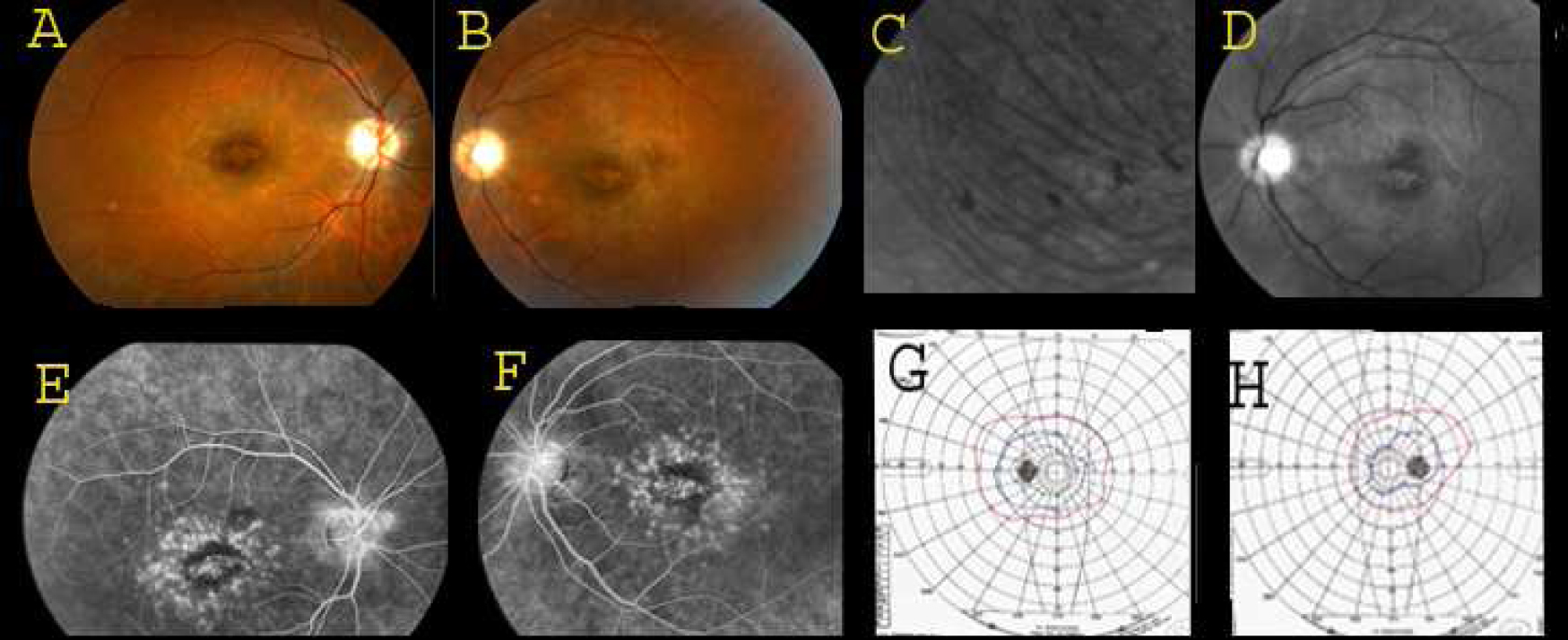
 Figure 2 of Pras, Mol Vis 2009; 15:1709-1716.