Figure 4. In vivo confocal microscopy (IVCM): Heidelberg Retina Tomograph II, Rostock Corneal Module images of individual II:3. A: IVCM image at the level of the basal epithelium, demonstrating two irregular amorphous regions of Bowman layer protruding
forwards into the epithelium, visible in the upper and lower aspect of the image. B: IVCM image at the level of Bowman layer, demonstrating a diffuse, irregular hyper-reflectivity. Sub-basal nerves are not
visible. C: IVCM image at the level of the anterior stroma, demonstrating patchy areas of hyper-reflectivity within the stroma with
hyper-reflectivity of keratocytes. D: IVCM image at the level of the midstroma, demonstrating unusual, tortuous stromal nerves with normal keratocytes. E: IVCM image at the level of the posterior stroma demonstrating normal architecture and normal keratocytes. F: IVCM image at the level of the endothelium, demonstrating normal healthy endothelium.
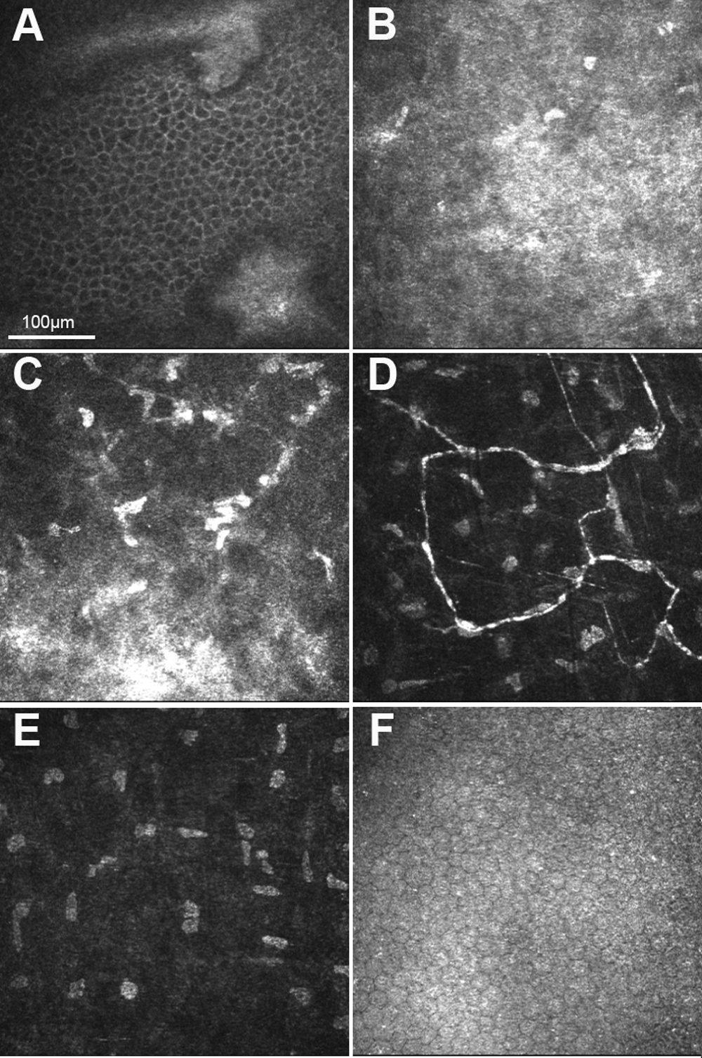
 Figure 4 of
Vincent, Mol Vis 2009; 15:1700-1708.
Figure 4 of
Vincent, Mol Vis 2009; 15:1700-1708.