Figure 3. In vivo confocal microscopy
(IVCM): Confoscan 2 images of I:2. A: IVCM image of normal
epithelium with prominent nuclei visible. B: IVCM image at the
level of Bowman membrane demonstrating abnormal diffuse
hyper-reflectivity. C: IVCM image at the level of the posterior
stroma demonstrating a normal keratocyte appearance with no evidence of
hyper-reflective structures (flecks) either within the keratocytes, or
in the extracellular regions. D: IVCM image at the level of the
endothelium, demonstrating normal healthy endothelium.
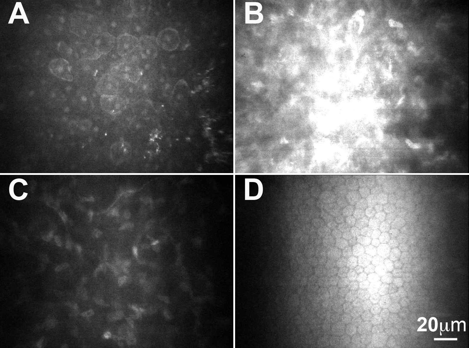
 Figure 3 of Vincent, Mol Vis 2009; 15:1700-1708.