Figure 2. Slit lamp images of corneas of
affected family members. Slit lamp images of cornea highlighting
diffuse, small, gray anterior stromal flecks (A-F) and larger,
discrete circular/oval/annular gray/white opacities (0.2–1.5 mm) at the
level of Bowman's layer (C-F, small arrowheads) and prominent
corneal nerves (D, fine arrow). A: Slit lamp image of
right cornea of individual I:2, age 64 years, at 10× magnification,
demonstrating diffuse small gray anterior stromal flecks and annular
opacities. B: Slit lamp image of left cornea of individual
II:5, age 37 years, at 10× magnification demonstrating diffuse small
gray anterior stromal flecks and annular opacities. C and D:
Slit lamp images of right cornea of individual II:3, at 10×
magnification (C) and 16× magnification (D). The annular
opacities are clearly visualised (small arrowhead) with prominent
corneal nerves visible (fine arrow; E and F) Slit lamp
images of right and left corneas respectively of individual III:2, age
14 years, at 10× magnification showing the annular opacities
highlighted with arrowheads.
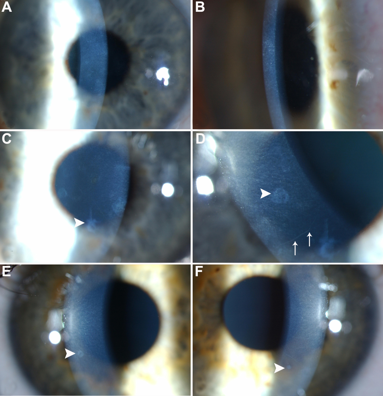
 Figure 2 of Vincent, Mol Vis 2009; 15:1700-1708.