Figure 4. Predominant expression of
Chop2-GFP in AII amacrine cells. A: In the retinal whole-mount,
GFP-positive AII amacrine cells were found throughout the retina. B,
C: At high magnification in the retinal whole-mount, GFP
expression was detected in the somas of AII cells in the INL (B)
and in the arboreal dendrites in the IPL (C). D: In the
retinal vertical section, many AII amacrine cells were GFP-positive. E-G:
In the same retina shown in D, GFP-positive cells (E)
were immunolabeled for Disabled-1 (F). The merge image is shown
in G. Double-labeled AII amacrine cells are marked by arrows.
For A-C, GFP fluorescence was enhanced with antibodies
against GFP. Abbreviations: outer nuclear layer (ONL); outer plexiform
layer (OPL); inner nuclear layer (INL); inner plexiform layer (IPL);
ganglion cell layer (GCL). Scale bars equal 500 μm in A, and 25
μm in B-D.
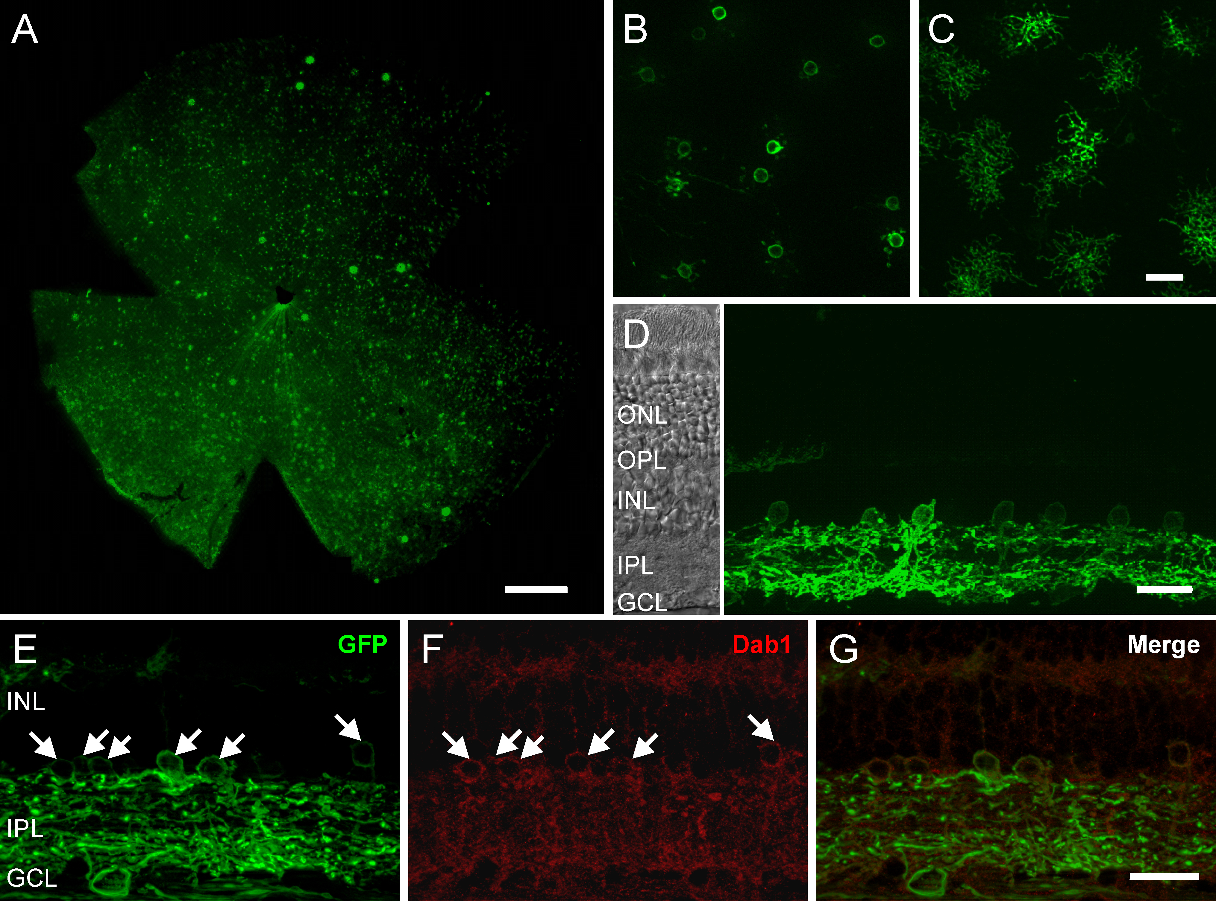
 Figure 4 of Ivanova, Mol Vis 2009; 15:1680-1689.