Figure 1. Representative fluorescent image
shows Chop2-GFP expression in retinal whole-mount preparation. Retinas
were photographed in 12 standard areas of 450×335 μm2
(dashed rectangles). The number of cells in the GCL was counted in
characteristic smaller areas of 0.04 mm2 shown by solid
rectangles (200×200 μm2).
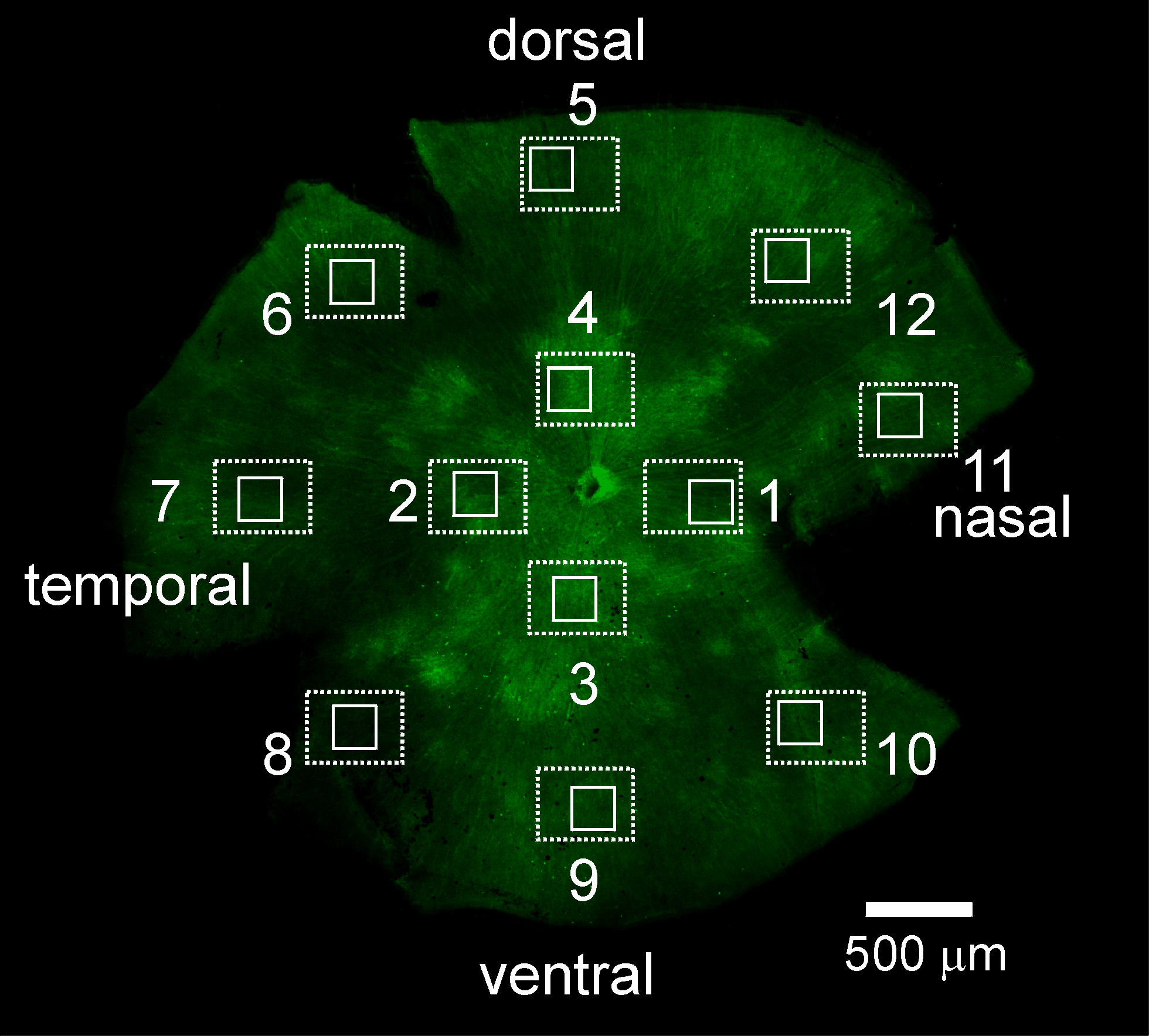
 Figure 1 of Ivanova, Mol Vis 2009; 15:1680-1689.