Figure 5. Shared morphological features of
rhodopsin-positive or PNA-positive cells in the pars plana and
peripheral retina in rd1 mice. Double-headed arrows indicate
the pars plana. A: At P30, many rhodopsin-positive cells in
both the ciliary epithelium and peripheral retina were oriented
randomly, while most of those in the posterior parts of the retina were
aligned radially in rd1 mice. B-D: Selected areas from A
are magnified. E: At P30, PNA-positive processes were generally
shorter in the peripheral retina and par plana compared to those in the
posterior parts of the retina with longer processes in rd1
mice. A and E are thick scans (merged from 4 scans;
each scan was 10.0 μm thick). Scale bar equals 50 μm. Abbreviations:
ciliary body (CB); recoverin (Rcv).
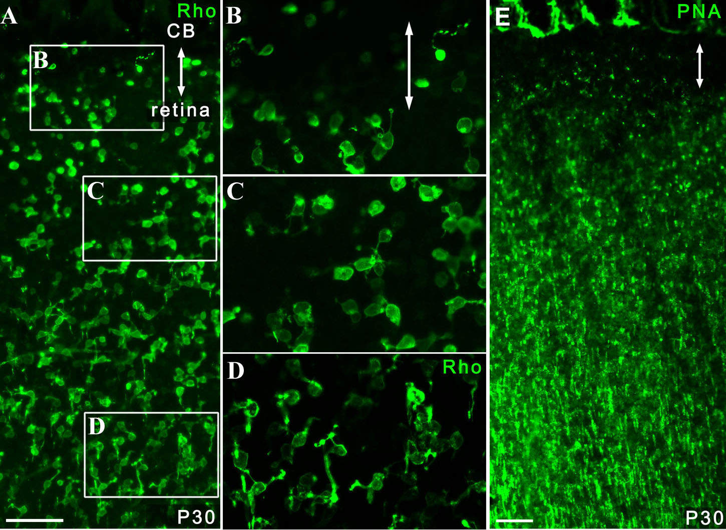
 Figure 5 of Nishiguchi, Mol Vis 2009; 15:187-199.