Figure 2. Identification of photoreceptor
lineage cells in the pars plana in P6 rd1 mice. Double-headed
arrows indicate the pars plana. All the images were obtained from P6 rd1
mice. A: Cells positive for both BrdU and Pax6 formed the
neuroblast layer that extended both in the peripheral retina and
ciliary body (n=6). B, C: The neuroblast layer composed of
BrdU-positive cells extended into the pars plana in which
rhodopsin-positive cells were identified (n=13). Note that a small
linear gap (arrowhead) demarcating the pars plana and retina can be
seen in B. C is a merged image of B and the
result of rhodopsin staining. D: Recoverin-positive cells were
observed in sparse distribution in the pars plana, while the peripheral
retina showed dense recoverin immunoreactivity (n=12). E, F:
The majority of recoverin-positive cells in the pars plana were cells
of cone photoreceptor lineage with a short PNA-positive process (n=7). F
is an enlarged image of E. Note that a linear gap (arrowhead)
demarcating the pars plana and retina can be seen. B, C, E, and
F are presented as a thin scan (a single scan 7.1 μm thick). D
is a thick scan merged from 2 images (each scan was 10.0 μm thick). A
dose of BrdU injected was 150 mg/kg. Scale bar equals 25 μm in D
and F and 50 μm in A and B. Abbreviations:
rhodopsin (Rho); recoverin (Rcv).
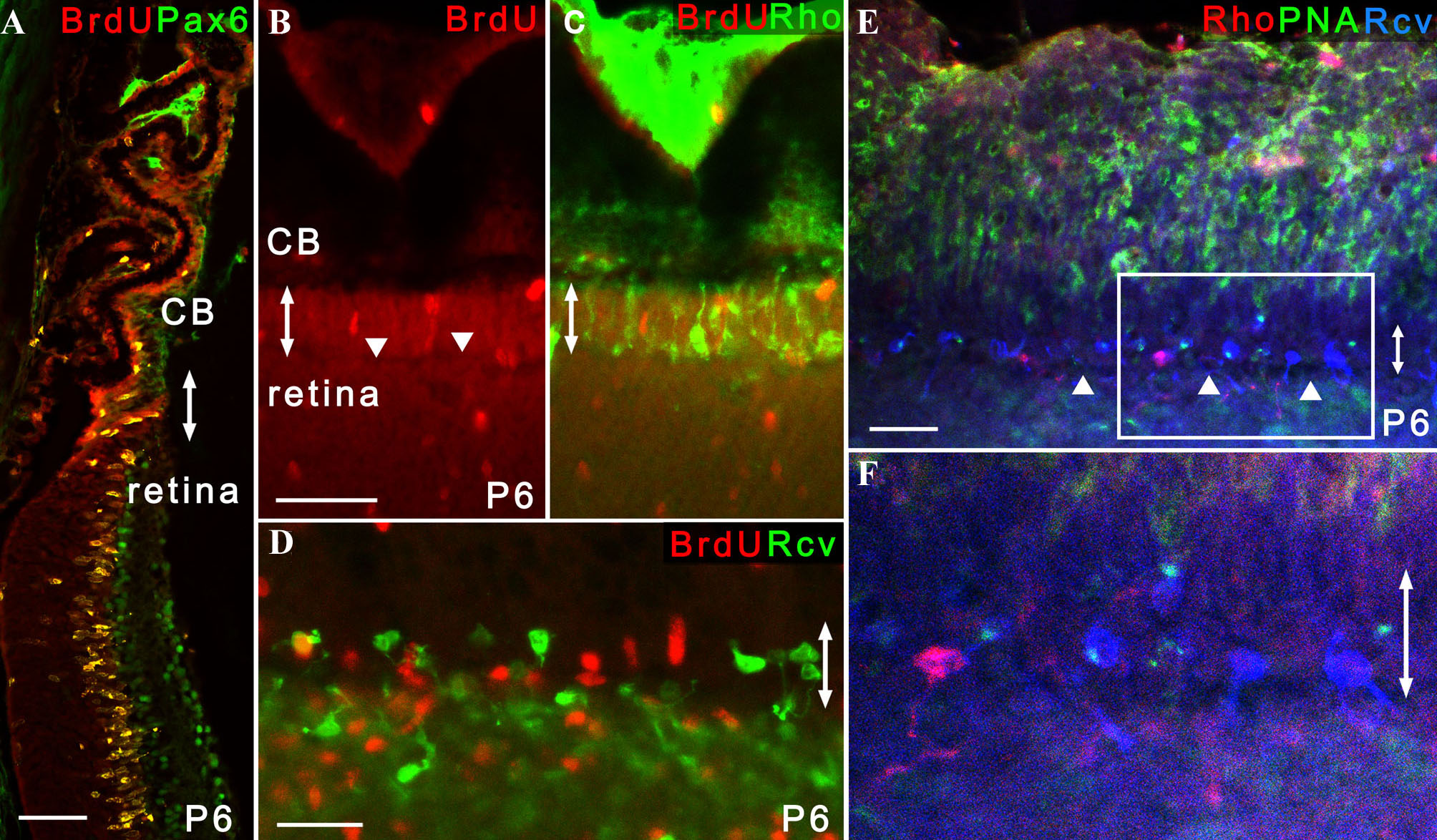
 Figure 2 of Nishiguchi, Mol Vis 2009; 15:187-199.