Figure 1. Cilioretinal flat-mounts. A: The diagram illustrates the histological section of the retina and ciliary body. Photoreceptors and their precursors are
shown as green cells in the deep retina and pars plana. B: Thin scans of the cilioretinal flat-mounts focused at the level of the ciliary epithelium showed cells positive for photoreceptor
markers in the pars plana as sporadic cells aligned circumferentially along the retinal margin. With this scanning method,
no photoreceptors could be seen in the retina. C: In thick scans of flat-mount specimens, dense photoreceptors in the deep retina were also visualized showing a sharp demarcation
at the cilioretinal border. Abbreviations: cornea (cor); ciliary body (cb).
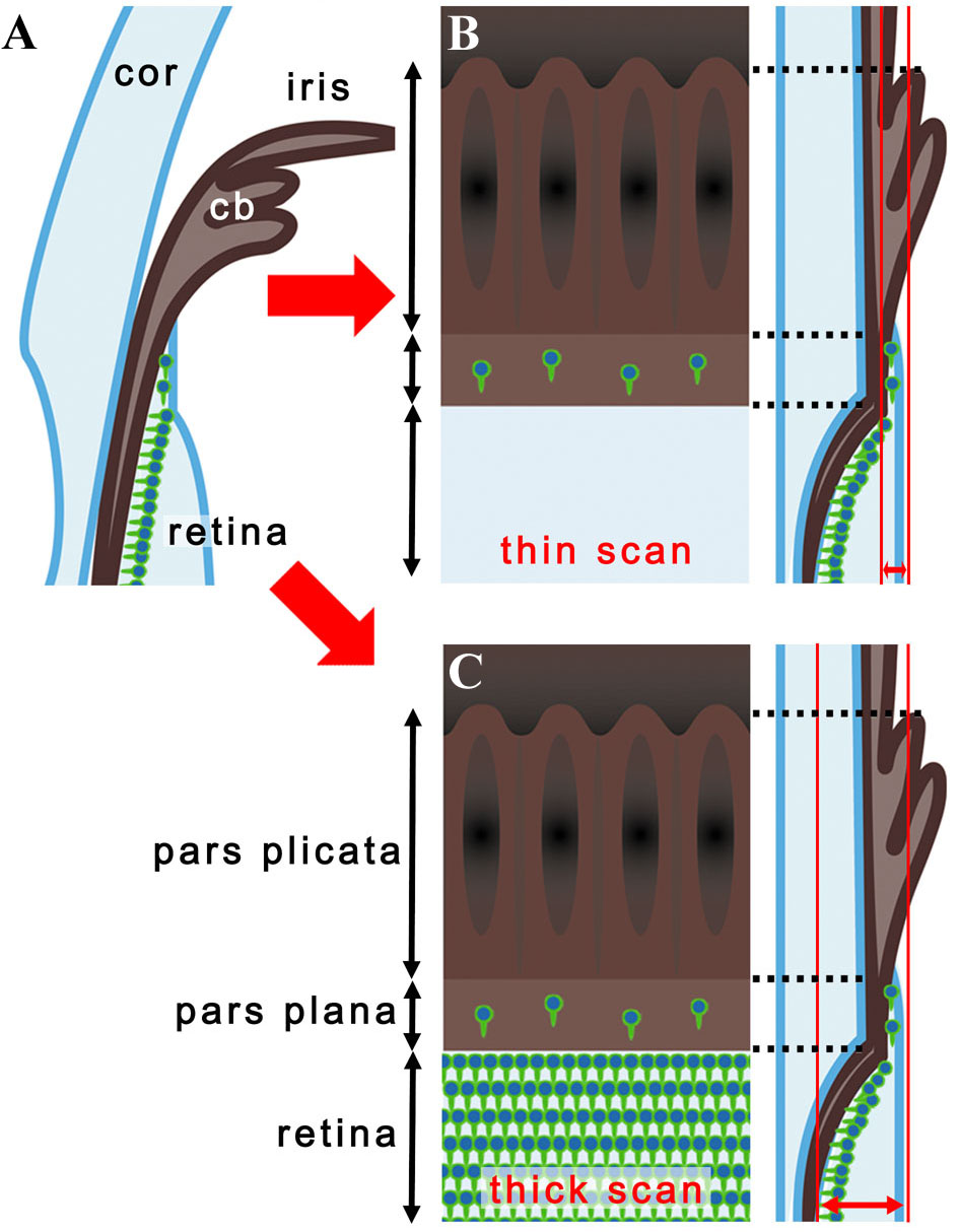
 Figure 1 of
Nishiguchi, Mol Vis 2009; 15:187-199.
Figure 1 of
Nishiguchi, Mol Vis 2009; 15:187-199.