Figure 1. Lack of LIF increases
light-induced photoreceptor degeneration. A: Retinal morphology
of Lif+/– and Lif–/– mice was
analyzed before (upper panels) or at 9 days after exposure to
15,000 lx of white light for 2 h (lower panels). Fewer
photoreceptors survived light exposure in the lower central retina (the
most affected region) in the absence of LIF in Lif–/–
mice. Shown are representative sections of at least three animals. B:
Rhodopsin levels were determined spectrophotometrically at 9 days after
light exposure as a quantitative assessment of surviving rod
photoreceptors in the whole retina. Rhodopsin levels after light
exposure were expressed relatively to the respective dark controls,
which were set to 100%. Note that values represent the average
rhodopsin content of the whole retina, whereas the morphological
pictures (A) show only the most affected areas. Abbreviations:
retinal pigment epithelium (RPE); rod outer segments (ROS); rod inner
segments (RIS); outer nuclear layer (ONL); inner nuclear layer (INL).
Number of animals (N) is indicated for each group. The asterisk
(*) indicates a p value of 0.0164 as calculated by a two-tailed t-test.
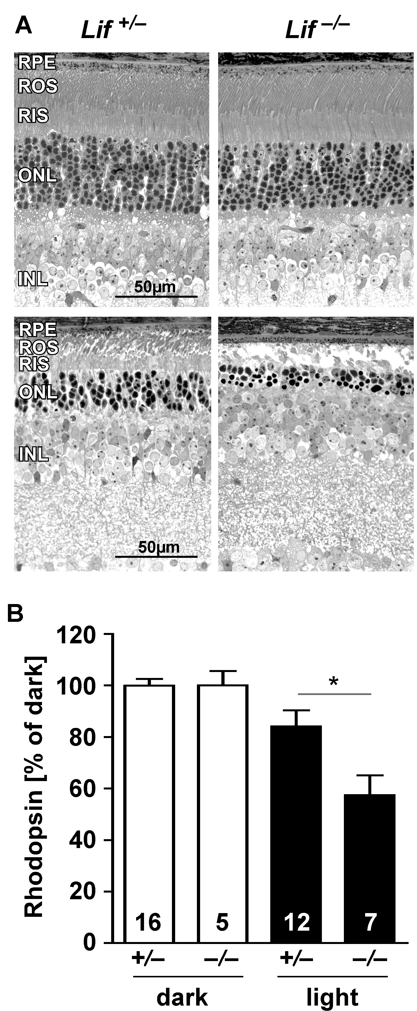
 Figure 1 of Bürgi, Mol Vis 2009; 15:1631-1637.