Figure 3. Diff-Quik staining photos and ex
vivo confocal microscopy scans of the brush cytology specimens from
patients with atopic keratoconjunctivitis. A and B:
Corresponding brush cytology photos and confocal microscopy scans from
a representative 12-year-old female patient. Confocal microscopy could
effectively discern the nuclear details such as segmentation in
polymorphs (yellow arrow heads), epithelial cell clumps (yellow
arrows), nuclei in conjunctival epithelial cells (which appeared as
round hyperreflective oval bodies) and the mucin blots appeared as
hyperreflective bodies with similar/exact shapes resembling the
Diff-Quik stained specimens (blue arrows). Corresponding brush cytology
photos and confocal microscopy scans from a representative 17 year old
male patient are also shown in C and D. Confocal
microscopy could effectively discern the nuclear details such as
segmentation in polymorphs (yellow arrow heads), and the double nuclei
in eosinophils (orange arrow heads) with similar/exact shapes
resembling the Diff-Quik stained specimens.
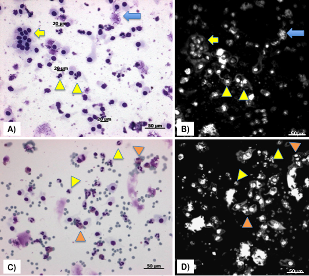
 Figure 3 of Wakamatsu, Mol Vis 2009; 15:1611-1619.