Figure 1. Conjunctival slit-lamp photograph, confocal microscopy images and brush cytology photos from a normal control subject and
an AKC patient. A: Slit lamp photograph of the upper tarsal conjunctiva in a 26-year-old male healthy control subject. Note the absence of
papillary formations. The conjunctival injection grade was 1 point. B: Representative confocal scan of the upper tarsal conjunctiva of the same subject. The inflammatory cell density was 256
cells/mm2. C: Representative photograph of the upper tarsal conjunctival brush cytology specimen of the same subject. The inflammatory
cell density was 320 cells/mm2. (Inflammatory cells shown by blue arrows). D: Slit lamp photograph of the upper tarsal conjunctiva in a 27-year-old male patient with AKC. Note the cobble stone like
papillary formations. The conjunctival injection grade was 3 points. E: Representative confocal scan of the upper tarsal conjunctiva of the same patient. The inflammatory cell density was 1,037
cells/mm2. F: Representative photograph of the upper tarsal conjunctival brush cytology specimen of the same patient. The inflammatory
cell density was 856 cells/mm2. Inflammatory cells were shown by blue arrows.
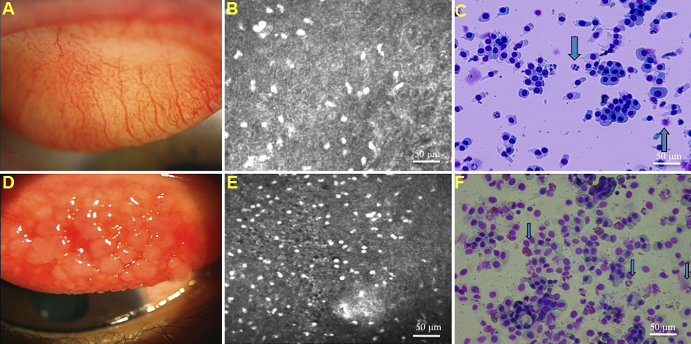
 Figure 1 of
Wakamatsu, Mol Vis 2009; 15:1611-1619.
Figure 1 of
Wakamatsu, Mol Vis 2009; 15:1611-1619.