Figure 1. Concentration dependency for
imatinib-induced phenotypic changes in RGC-5 cells. Subconfluent RGC-5
cells were maintained in complete medium (+FBS) or serum-free medium
(-FBS) for 48 h, during which time the cells were exposed to increasing
concentrations of imatinib (1 µM to 30 µM; A, B). In addition,
serum-free medium (-FBS) was supplemented with either 30 ng/ml PDGF or
30 nM insulin during RGC-5 cell exposure to imatinib (C, D). The
apoptotic phenotype was then assessed by nuclear DAPI staining
(fluorescence microscopy) and morphological changes (phase-contrast
microscopy). The images shown are representative of 4 to 5 separate
experiments.
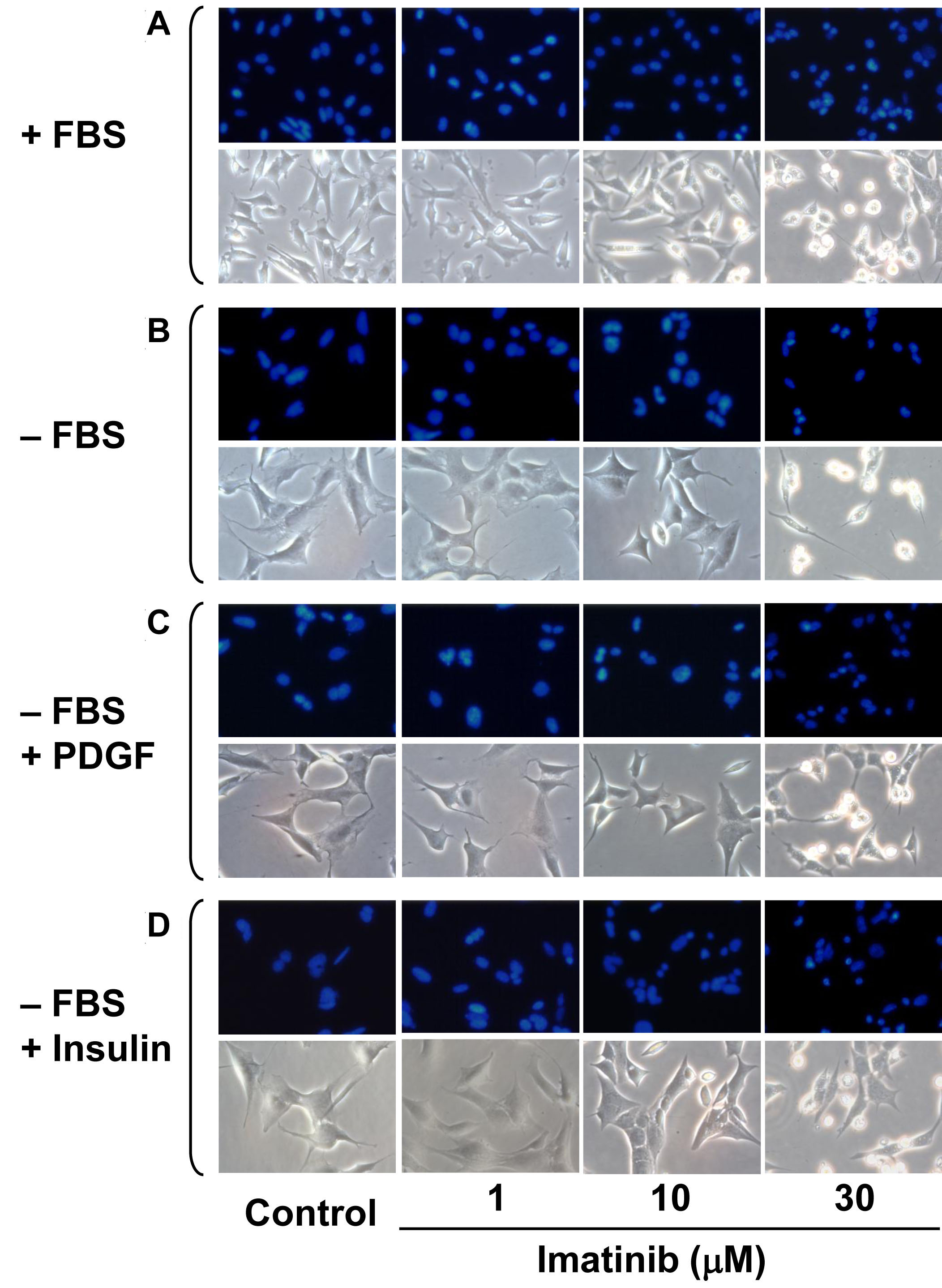
 Figure 1 of Biswas, Mol Vis 2009; 15:1599-1610.