Figure 4. Immunohistochemical staining of MCEC sheets. Paraffin sections (5 µm) were stained with; A = H&E, B = immunostained with anti-cytokeratin 12, and C = normal goat IgG. Normal mouse cornea was immunostained with anti-cytokeratin 12 (D). Scale bar; 50 µm.
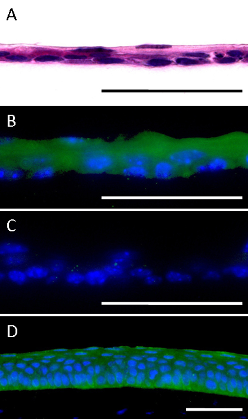
 Figure 4 of
Kobayashi, Mol Vis 2009; 15:1589-1593.
Figure 4 of
Kobayashi, Mol Vis 2009; 15:1589-1593.