Figure 4. Confocal microscopy analysis of the proband’s left cornea. Superficial epithelial cells (A, B), wing cells (C, D), Bowman’s membrane (E,F), anterior keratocytes (G, H), and posterior keratocytes (I, J) on GDLD (A, C, E, G, I) and normal cornea (B, D, F, H, J) are displayed. A: The epithelial cells of GDLD cornea were irregular in shape and often elongated. B: Normal epithelial cells are shown. C: The overall epithelial architecture was destroyed. D: Normal wing cells are shown for comparison. E: At the level of the Bowman’s membrane, a very small number of sub-basal nerves were detectable. F: Normal Bowman’s membrane is shown. G: Large accumulations of brightly reflective amyloid materials were noted within the anterior stroma. H: Normal anterior keratocytes are shown for comparison. I: Posterior keratocytes of the GDLD patient. J: Posterior keratocytes of a normal control.
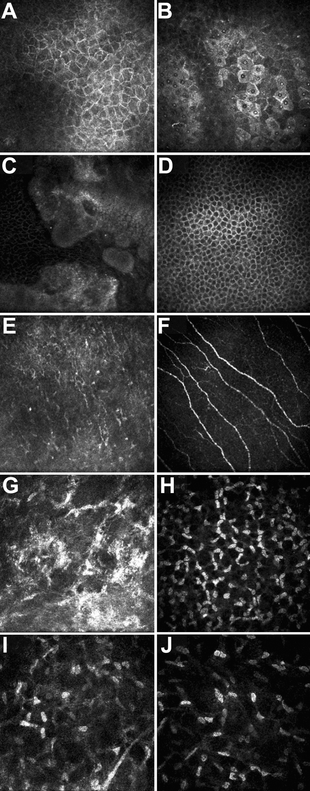
 Figure 4 of
Jing, Mol Vis 2009; 15:1580-1588.
Figure 4 of
Jing, Mol Vis 2009; 15:1580-1588.