Figure 3. Photography of corneal
histological features of the proband. A: A hematoxylin and
eosin stained section shows epithelial atrophy, epithelial hyperplasia,
and the considerable extent of the subepithelial and superficial
stromal deposits of eosinophilic material. Original magnification 100X.
B: Nodular subepithelial amorphous deposits stain positive with
periodic acid-Schiff. Original magnification 100X. C: Congo
red-positive accumulations were within or beneath the epithelium and
within the anterior stroma. In some areas, Congo red-positive deposits
accumulated to form gelatinous drop-like masses. Original magnification
100X. D: Higher magnification revealed band-like structures
with a spike-like pattern just beneath the epithelium. Original
magnification 400X. E: Congo red staining under polarized light
revealed typical apple-green birefringence in epithelial and
subepithelial regions, indicative of amyloid. Original magnification
100X.
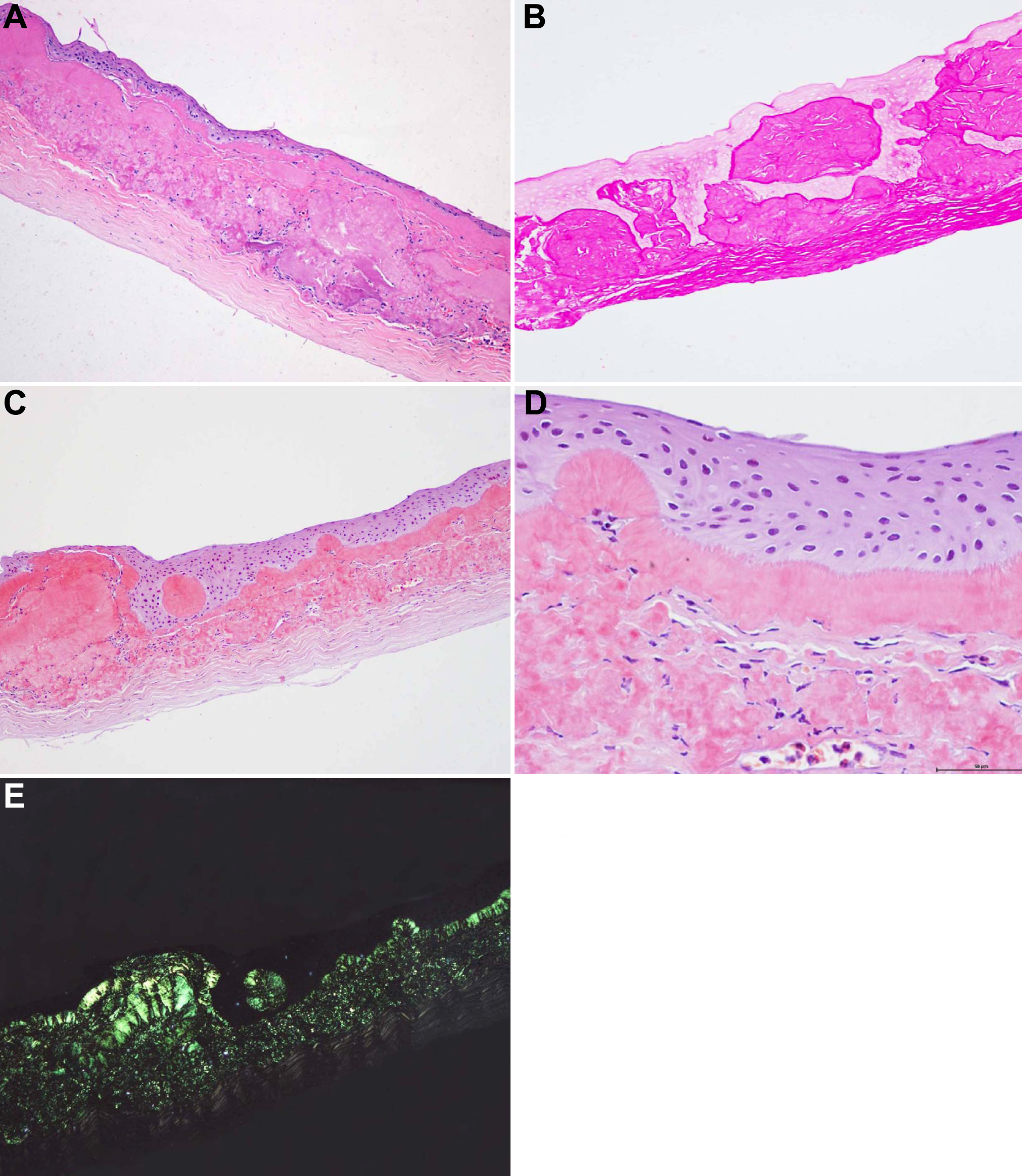
 Figure 3 of Jing, Mol Vis 2009; 15:1580-1588.