Figure 4. Histological findings in the rabbit eye with induced uveitis at 10 days after rAAV-IL-1Ra administration. The vitreal cavity
of rabbit eyes was injected with rAAV-IL1-Ra, and the contralateral eye was injected with the same amount of rAAV-LacZ. In
another group, the right eye was injected with the same amount of PBS. After 10 days, uveitis was induced by intravitreal
injection of IL-1α. The eyeballs were enucleated, fixed, and stained with H & E immediately before, or one or three days after,
uveitis induction. In the eyes injected with rAAV-LacZ, the time points are immediately before (A), one day after (B), and three days after (C) uveitis induction. In the eyes injected with PBS, the time points are immediately before (D), one day after (E) and three days after (F) uveitis induction. In the eyes injected with rAAV-IL-1Ra, the time points are immediately before (G), one day after (H), and three days after (I) IL-1α injection. The scale bar in I is equal to 100 μm.
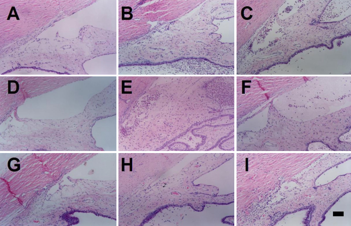
 Figure 4 of
Tsai, Mol Vis 2009; 15:1542-1552.
Figure 4 of
Tsai, Mol Vis 2009; 15:1542-1552.