Figure 1. Establishment of experimental uveitis. The vitreal cavity of rabbit eyes was injected with IL-1α to induce experimental uveitis,
and the contralateral eye was injected with the same amount of the vehicle (distilled water) as a control. Inflammation was
evaluated by the presence of leukocyte infiltration (arrowheads) in the tissues of the anterior ocular segment. Immediately
before IL-1α or vehicle injection, no leukocyte infiltration was observed (A, B). The inflammation, defined as the presence of massive leukocyte infiltration in the tissue of the anterior ocular segment,
peaked one day after IL-1α injection (C). Three days after IL-1α injection, the inflammation had partially subsided (E), and 15 days after injection, no sign of inflammation was seen in the anterior ocular segment (G). Experimental uveitis was not detected in the control eyes at 1, 5, or 15 days after vehicle injection in the control group
(B, D, F, H). The scale bar in H is equal to 100 μm.
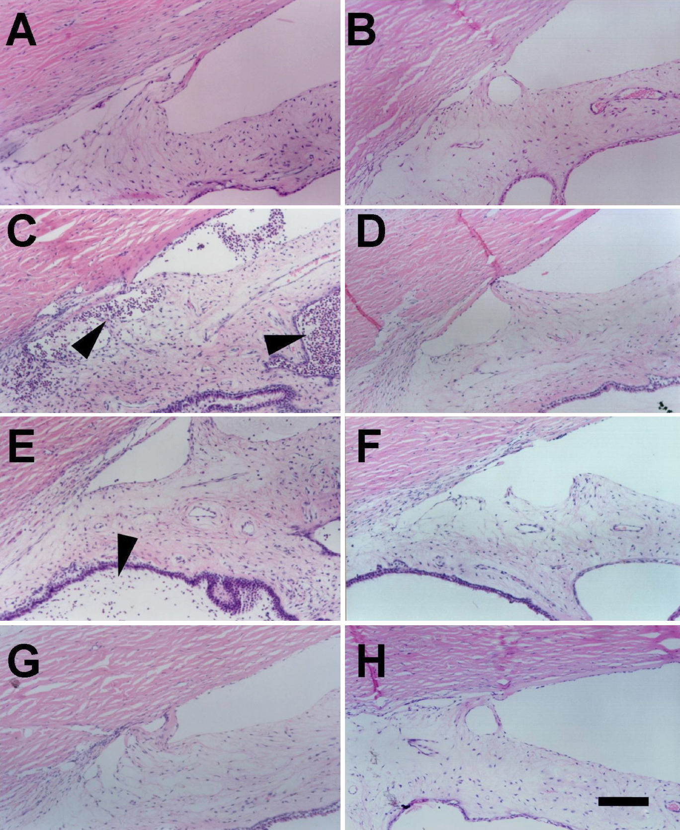
 Figure 1 of
Tsai, Mol Vis 2009; 15:1542-1552.
Figure 1 of
Tsai, Mol Vis 2009; 15:1542-1552.