Figure 2. AGE-modification of FN alters
integrin-mediated signaling in RMECs. A: western blotting
analysis showed that phospho-FAK (tyrosine397) was reduced
when RMECs were grown on AGE-FN. Quantification revealed this clear
differential, which is especially evident at the 45 and 60 min time
point. B-G: Confocal microscopy disclosed integrin α5β1
immunoreactivity in RMECs grown on FN and AGE-FN. B-D: RMECs
cultured on native FN exhibited a relatively uniform distribution of
α5β1 throughout the basal aspect of the cell immediately adjacent to
the substrate. Deeper (basal) z-sections showed relatively weak
fluorescence intensity at the basal plasma membrane (basal PM; D).
E-G: RMECs cultured on AGE-FN exhibited a greater intensity of
green fluorescence indicative of higher α5β1 expression. Some RMECs
exposed to AGE-FN showed perinuclear punctuate staining (F).
Deeper z-sections showed high fluorescence intensity and a filamentous
distribution at the basal PM (G). All cells were counterstained
with propidium iodide.
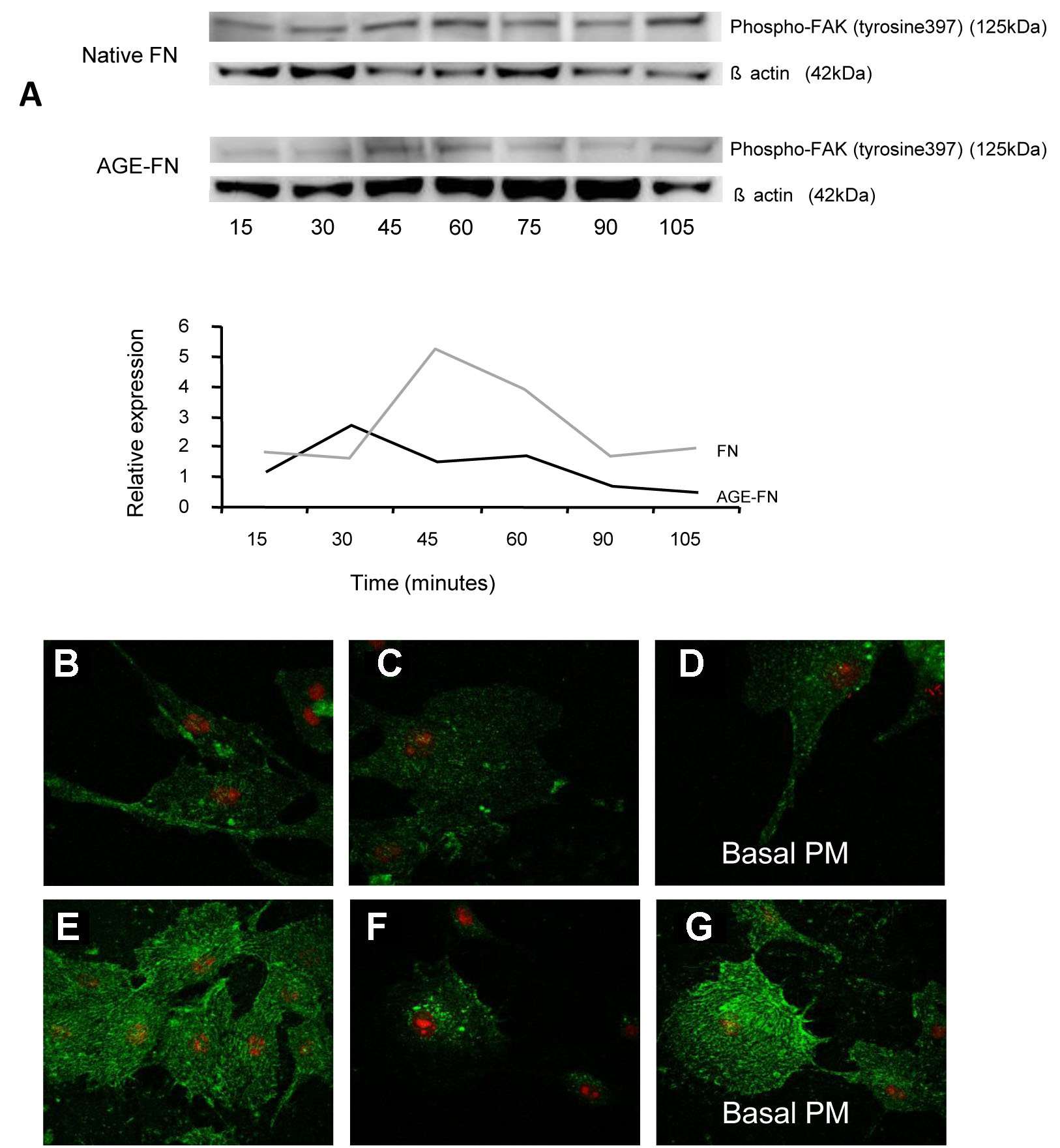
 Figure 2 of McDonald, Mol Vis 2009; 15:1509-1520.