Figure 8. Filipin-treated gap junctions. A:
A representative cholesterol-rich gap junction with the presence of
numerous filipin-cholesterol complexes (FCCs, small arrows) is
frequently found in the outer cortical fibers. B and C:
Cholesterol-intermediate gap junctions with less number of FCCs (small
arrows) are observed in the inner cortical fibers. FCCs (small arrows)
are often distributed along patches or parallel rows of
crystalline-packed connexons (large arrows). D and E:
Cholesterol-free gap junctions are mostly seen in the deeper mature
cortical fibers. Note that crystalline-arranged connexons can be
visualized more clearly on the E-face (ef) of the membrane. In the
images, pf indicates the P-face of the membrane. The scale bars
indicate 200 nm.
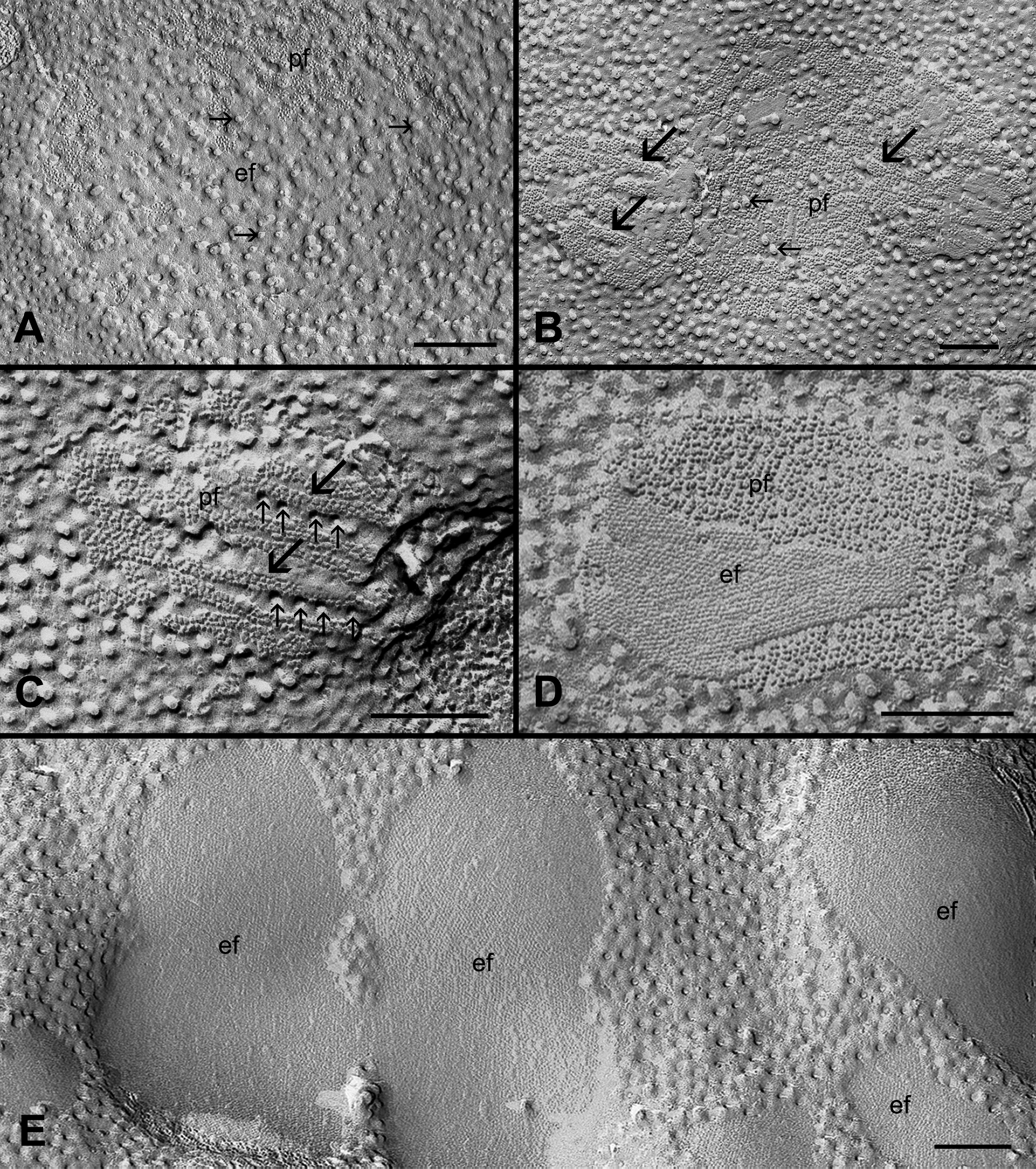
 Figure 8 of Biswas, Mol Vis 2009; 15:1492-1508.