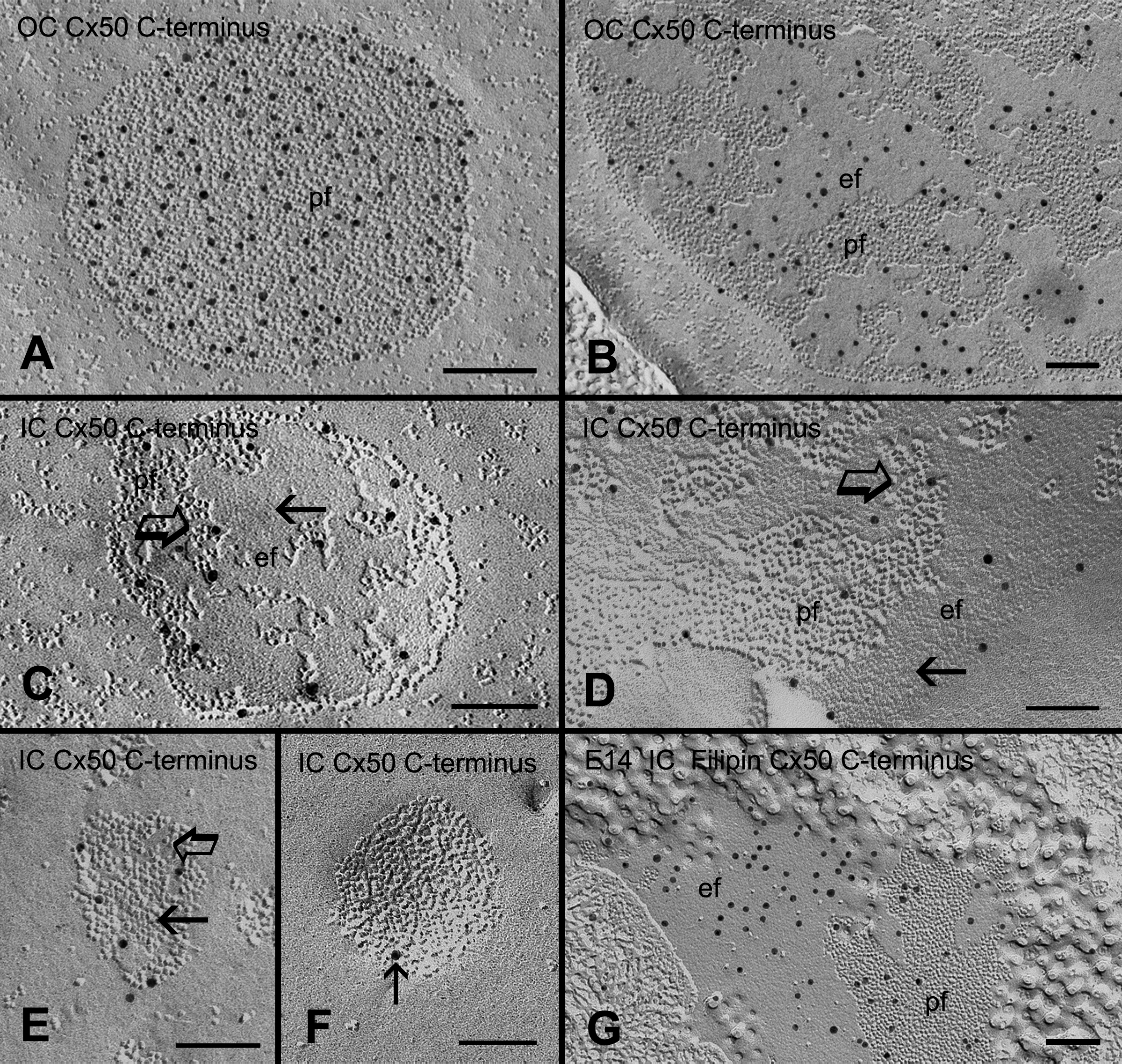
Figure 10. Freeze-fracture immunogold
labeling (FRIL) of the Cx50 COOH-terminus in different gap junction
configurations. FRIL showing the specific labeling of the COOH-terminus
of chick Cx50 antibody in gap junctions in the outer cortex (
A,
B)
and in the inner cortex (
C-
F) at high magnification. In
the young outer cortical fibers (OC), immunogold labeling of the Cx50
COOH-terminus antibody can be observed specifically on the P-face (
A)
and E-face (
B) of gap junctions with loosely-packed connexons.
However, a considerably smaller number of immunogold labeling
(particle) of the Cx50 COOH-terminus antibody is seen in gap junctions
with a mixture of crystalline-packed (arrows) and loosely-arranged
(open arrows) connexons in the inner cortex (
C-
F). Note
that only a single immunogold particle (arrow) is seen in this GJ with
crystalline-packed connexons in (
F). For comparison, in the
embryonic chick lens (
G) in which the gap junctions do not
display the distinct crystalline-packed connexons [
7], many immunogold
particles can be observed in the gap junction with cholesterol-free and
non-crystalline-packed connexons. The scale bars indicate 100 nm.
 Figure 10 of Biswas, Mol Vis 2009; 15:1492-1508.