Figure 4. CERKL-HA (isoform a, 532 amino
acids) shows a dynamic subcellular localization in COS-7 transfected
cells. A: Several cells per field showed uniform distribution
of CERKL in the cytosol and the nucleus, with clear exclusion from the
nucleoli. B: A similar pattern but with strong accumulation in
the nucleoli (nuclei, counter-stained with DAPI, appear in blue). C
and D: In most cells, CERKL was absent from the nucleus and
instead accumulated in clusters, preferentially in the perinuclear
region (A). These two patterns were cell-specific and could be
observed in the same field (D). E: CERKL localized to
several membranous subcellular compartments, mainly ER and Golgi. The
markers used were calnexin for ER, GM130 for Golgi, EEA1 for endosomes
and MitoTracker for mitochondria. F: R257X truncated CERKL
localized preferentially in the nuclei, although it was also detected
in the ER. Arrows highlight the relevant CERKL localizations, as
immunodetected with an anti-HA monoclonal antibody. Scale bar
corresponds to 10 μm. The same results were obtained for all the CERKL
isoforms, irrespective of the epitope used, HA or GFP (data not shown).
Abbreviations: endoplasmic reticulum (ER), endosomes (End),
mitochondria (Mit).
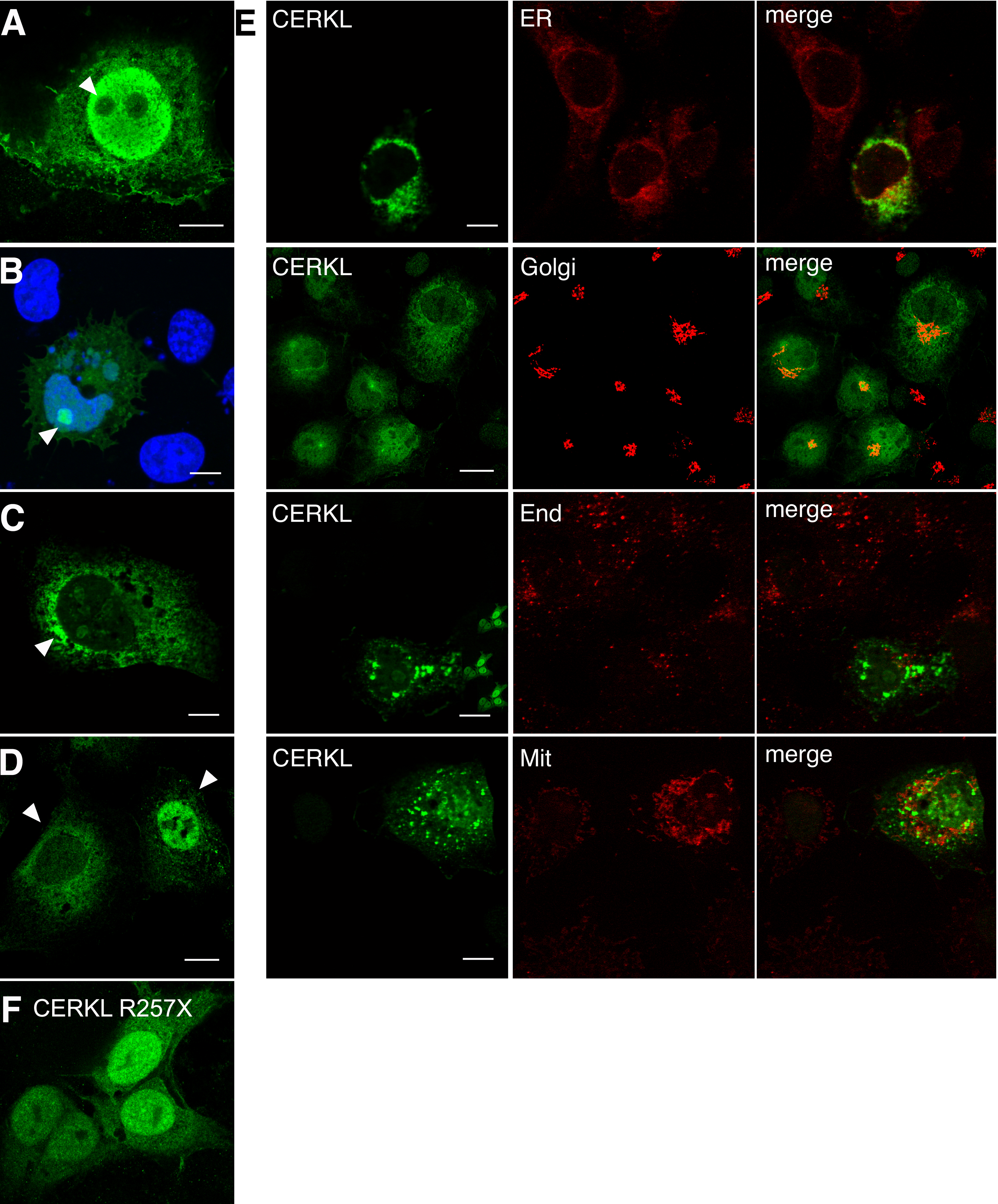
 Figure 4 of Tuson, Mol Vis 2009; 15:168-180.