Figure 3. CERKL kinase activity assays. A:
The autoradiography of one out of many thin layer chromatographies
(TLC) from in vitro assays is shown. Lipid micelles and
heat-inactivated cell lysates from 661W (murine photoreceptor-derived
cell line) cells were used as substrates, whereas protein lysates from
COS-7 cells transfected with either empty vector (pcDNA), CERKL isoform
a (532 aa), CERKL isoform b (558 aa) or ceramide kinase CERK (positive
control) expressing constructs were the source of the enzymatic
activity. B: Autoradiography of a TLC from in vivo assays is
shown. Cultured 661W cells were transfected with either pGFP (empty
vector), or CERKLa-GFP, CERKLb-GFP, or CERK-GFP (positive control),
selected by FACS and grown in a medium supplemented with 32P-orthophosphate
(see Material and Methods for details on the protocols). C1P represents
Ceramide-1-phosphate. The images are representative of many different
assays, with several experimental parameters, such as the type of cell
line and the CERKL isoforms overexpressed, changed. The results of the
assays were negative under all the conditions tested.
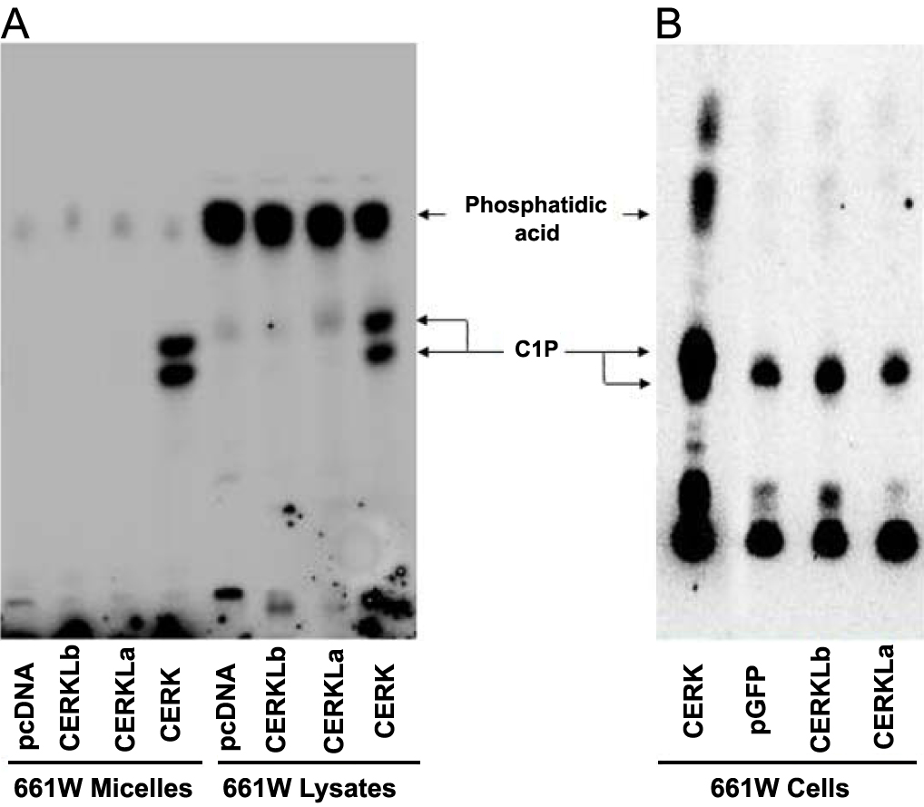
 Figure 3 of Tuson, Mol Vis 2009; 15:168-180.