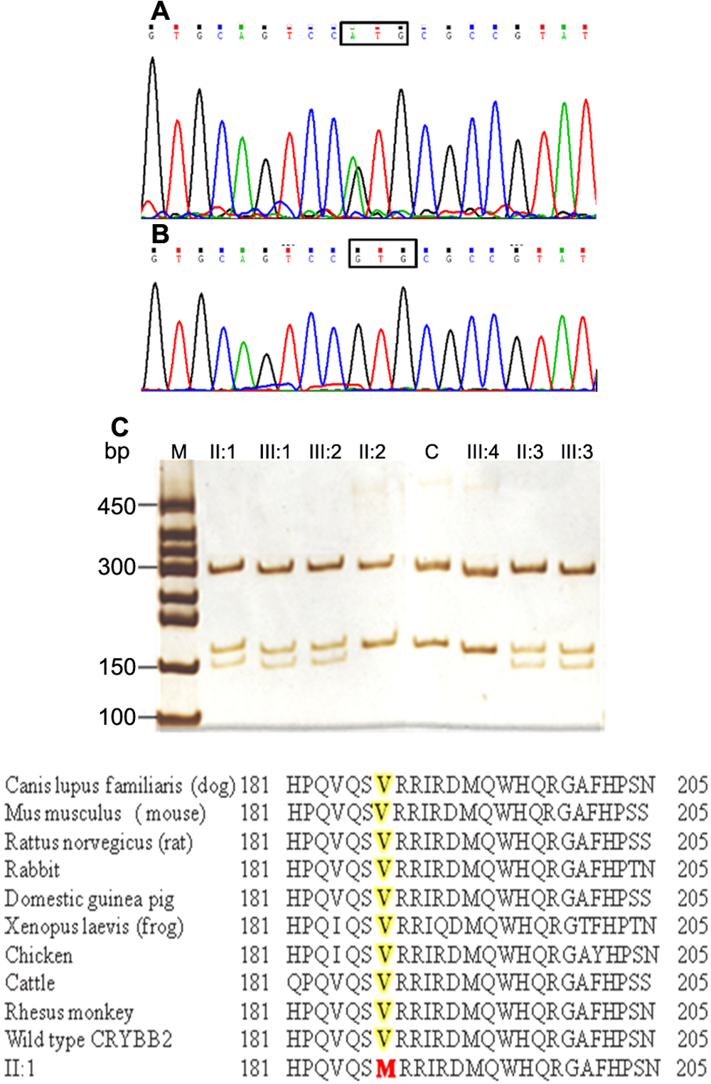
Figure 2. Mutation analysis of CRYBB2. The sequence chromatogram of a mutant allele shows a heterozygous G→A transition that changed Valine at codon 187 to Methionine
(counting the A of the ATG start codon as number 1; A). The sequence chromatogram of a wild type allele shows Valine (GTG) at codon 187(B). Restriction fragment length analysis showing that a gain of the novel NIaIII site cosegregated with affected individuals
heterozygous with the V187M mutation (300, 168, and 150 bp) but not with unaffected individuals (300 and 168 bp). C demonstrates an unrelated healthy individual. D: Sequences producing specific alignment of CRYBB2 amino acids. A protein–protein BLAST search (NCBI) of human βB2-crystallin
amino acid sequence was done. Proteins having various levels of sequence identity were picked and manually aligned. Shaded
letters (yellow) correspond to amino acid that is mutated in this study (highlighted in red).

 Figure 2 of
Mothobi, Mol Vis 2009; 15:1470-1475.
  Figure 2 of
Mothobi, Mol Vis 2009; 15:1470-1475.