Figure 3. In vivo laser scanning confocal
microscopic findings for the proband. A, B: Brightly reflective
deposition, presumably cholesterol or lipid, is associated with the
subepithelial nerve fibers in the left cornea. The shapes of the
crystals were needle-shaped or rectangular. C: Anterior stroma
showed the presence of multiple deposits of brightly reflective
crystalline material. The keratocyte nuclei are undetectable.
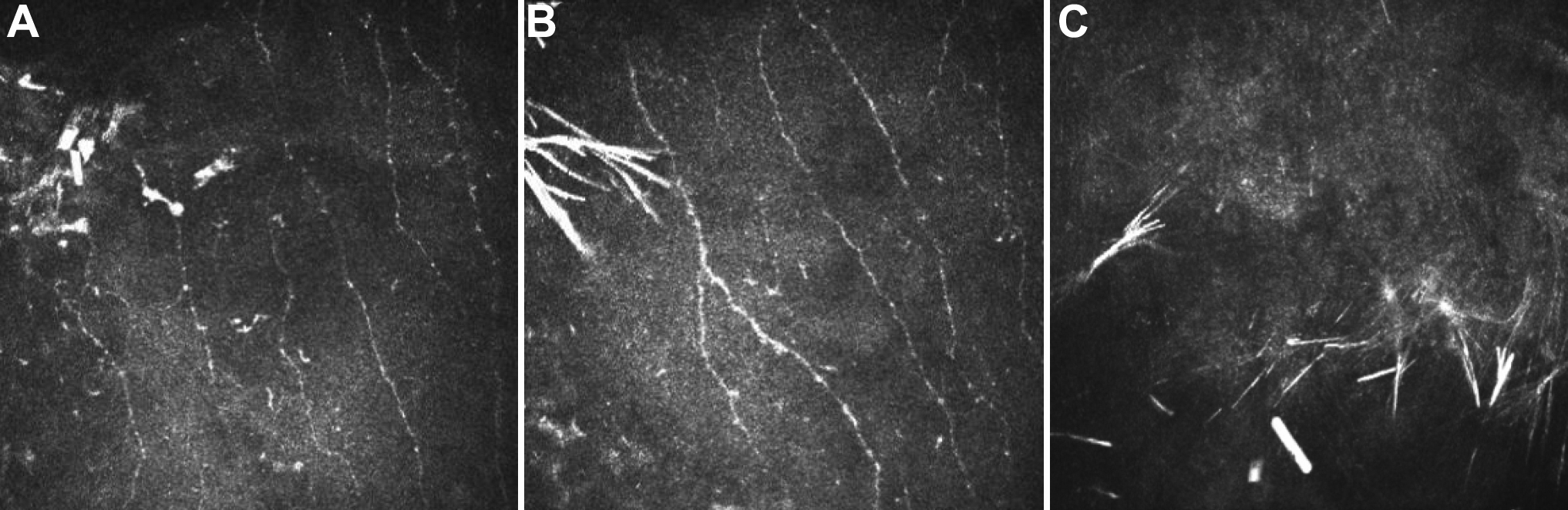
 Figure 3 of Jing, Mol Vis 2009; 15:1463-1469.