Figure 1. Slit-lamp photographs of
affected patients eyes. A: The right cornea of the 29-year-old
Chinese woman shows punctiform deposition of subepithelial crystals,
central haze, and arcus lipoides. B: Appearance of her left
cornea demonstrates a central plaque of subepithelial crystals slightly
inferiorly displaced in the visual axis, midperipheral clouding, and
arcus lipoides. C: Slit-lamp photograph of left eye (OS)
demonstrating subepithelial crystalline deposits. D: The right
cornea of the 54-year-old man shows central panstromal disc-like
opacity obscuring the pupillary axis, and prominent arcus lipoids. E,
F: Anterior stromal disciform opacity with arcus lipoides is
observed in his left eye. No crystals are apparent in the corneal
stroma.
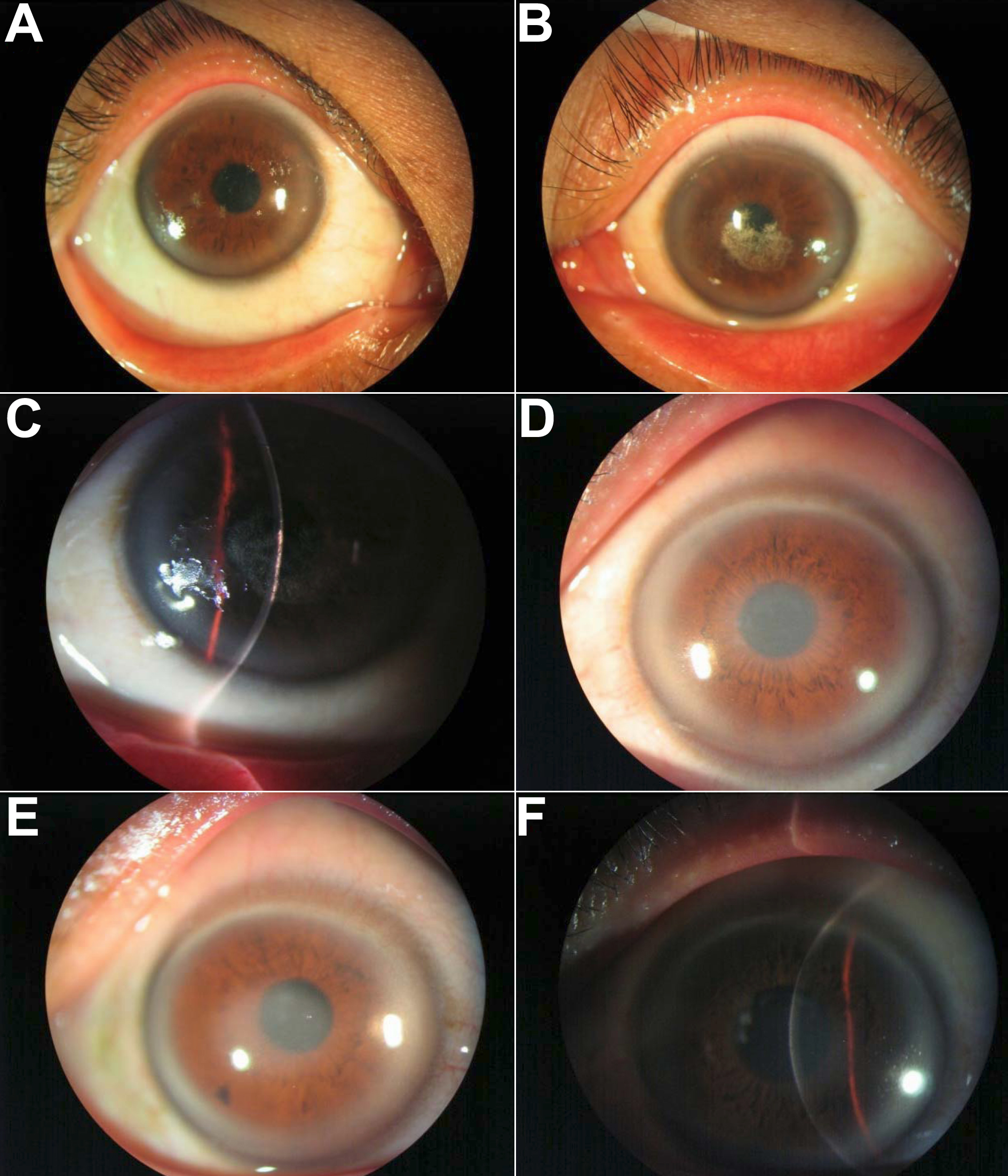
 Figure 1 of Jing, Mol Vis 2009; 15:1463-1469.