Figure 4. Representative HRTII-RCM images of the right cornea of patient #3 before and after treatment. The dotlike opacities (arrow)
and dendritic cells (arrow head) apparent in the basal cell layer of the corneal epithelium (BC) or in association with the
subepithelial nerve plexus (NP) before treatment were no longer evident after treatment. Scale bar, 50 µm.
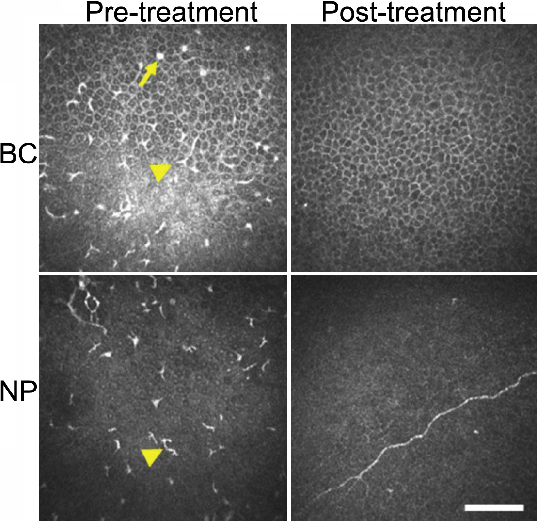
 Figure 4 of
Kawamoto, Mol Vis 2009; 15:1456-1462.
Figure 4 of
Kawamoto, Mol Vis 2009; 15:1456-1462.