Figure 3. Representative HRTII-RCM images of the subepithelial nerve plexus of the cornea in three patients with TSPK. Aggregated dendritic
cells were apparent in association with the nerve plexus (arrow) in both eyes of patient #1, in the left eye of patient #2,
and in the right eye of patient #3. Scale bar, 50 μm.
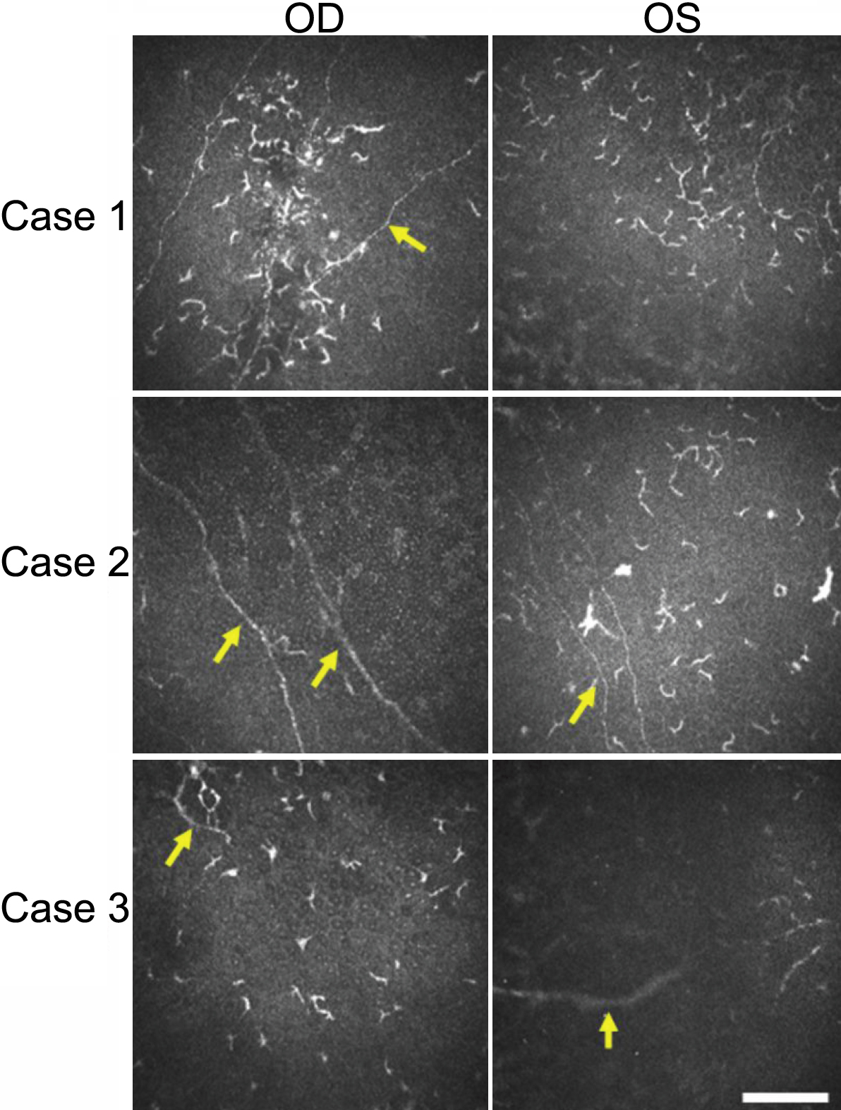
 Figure 3 of
Kawamoto, Mol Vis 2009; 15:1456-1462.
Figure 3 of
Kawamoto, Mol Vis 2009; 15:1456-1462.