Figure 2. Representative HRTII-RCM images of the basal cell layer of the corneal epithelium in three patients with TSPK. All images
show the honeycomb-like appearance characteristic of the normal cornea. Highly refractive dots and aggregation of dendritic
cells (arrow) were also evident in both eyes of patient #1, in the left eye (OS) of patient #2, and in the right eye (OD)
of patient #3. Scale bar, 50 μm.
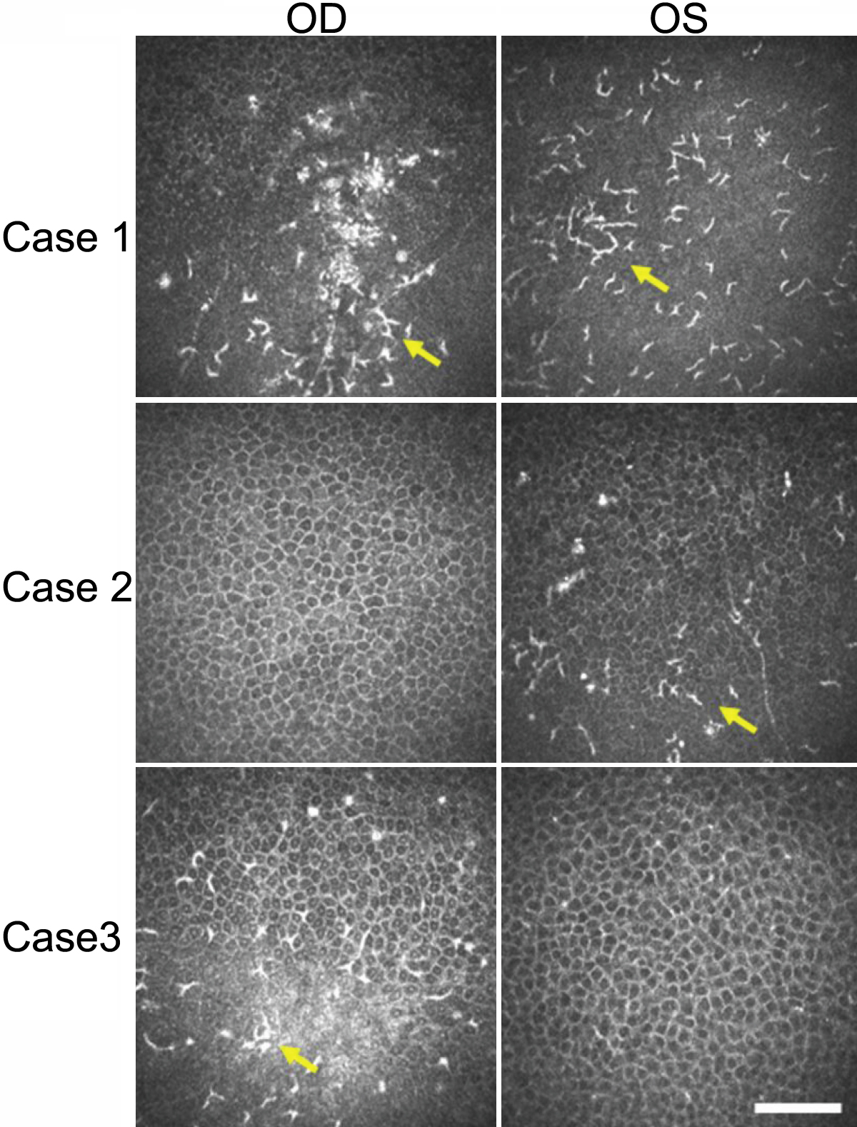
 Figure 2 of
Kawamoto, Mol Vis 2009; 15:1456-1462.
Figure 2 of
Kawamoto, Mol Vis 2009; 15:1456-1462.