Figure 1 of
Kawamoto, Mol Vis 2009; 15:1456-1462.
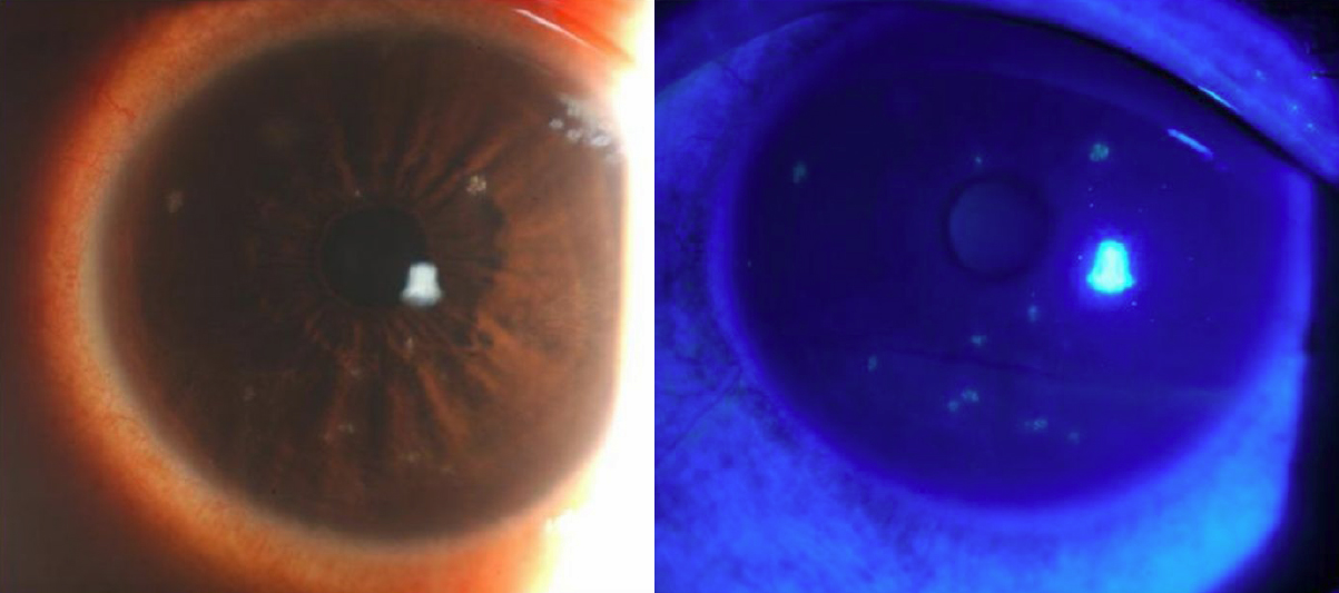
Figure 1.
Representative photographs of the affected eyes with TSPK. In the left eye of patient #1, multiple coarse granular opacities stained with fluorescein are seen in the cornea.
 Figure 1 of
Kawamoto, Mol Vis 2009; 15:1456-1462.
Figure 1 of
Kawamoto, Mol Vis 2009; 15:1456-1462.