Figure 2. Immunolocalization of Th17 cells
in OCP conjunctiva. Th17 lymphocytes were labeled with PE-conjugated
anti-human IL-17 antibodies (red). Increased Th17 staining was observed
in OCP (A) compared to healthy subjects (B).
Double-staining for CD4 (C, blue) and IL17 (D, red)
performed on four stage-III OCP samples demonstrated that 72% of CD4+ T
cells were Th17 lymphocytes (Merged image E, violet).
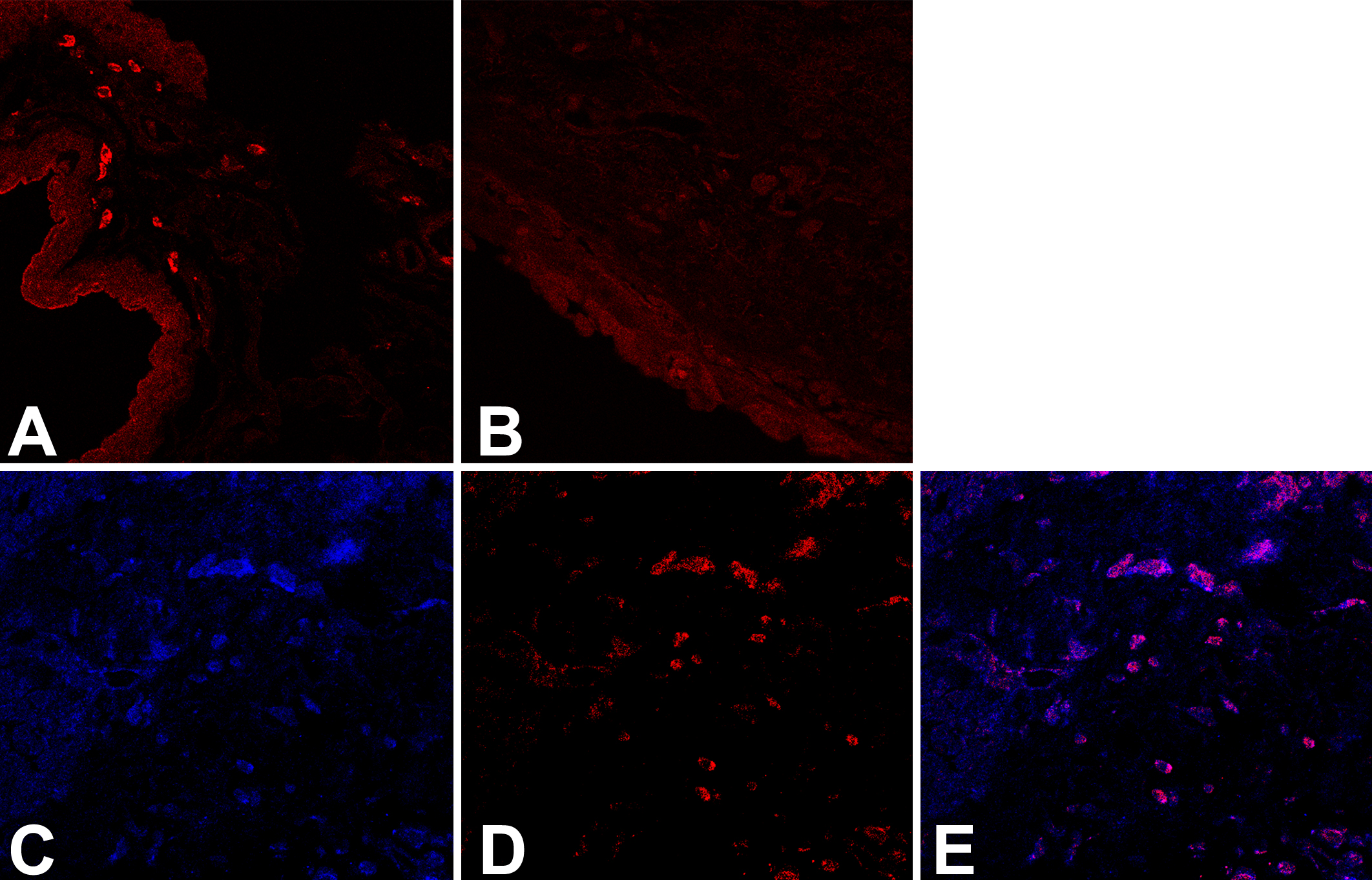
 Figure 2 of Lambiase, Mol Vis 2009; 15:1449-1455.