Figure 1. Histology of OCP conjunctiva.
OCP conjunctiva was infiltrated with immune cells, including
lymphocytes, plasma cells and leukocytes (A), whereas healthy
conjunctiva was free of immune cells (B). A linear direct
immunofluorescence labeling (green) of autoantibodies in the
conjunctival basal membrane was observed in all OCP samples (insert A).
No immunostaining was observed in control samples (insert B).
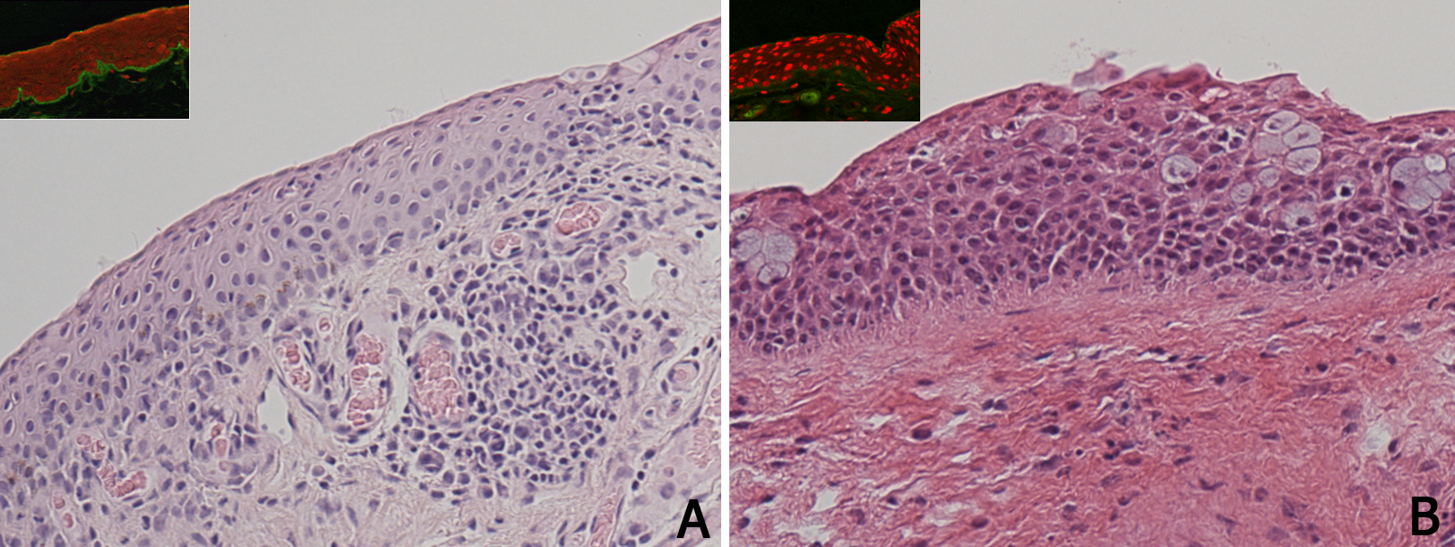
 Figure 1 of Lambiase, Mol Vis 2009; 15:1449-1455.