Figure 3. Isoelectric focusing of purified wild-type αB-crystallin and its mutants under native conditions. The approximate pI of isoelectric focusing gel was estimated from a pI calibration kit (pI range 3-10). The arrow indicated the position of bovine carbonic anhydrase B with a pI of about 5.85. A: Lane 1, WT; Lane 2, Δ2; Lane 3, Δ5; Lane 4, Δ7; and Lane 5, Δ10. B: Lane 1, Δ1; Lane 2, Δ1/K174A; Lane 3, Δ1/K174S; Lane 4, Δ1/K174E; Lane 5, K174A; Lane 6, K175A; Lane 7, K174/175A; Lane
8, K174/175E; and Lane 9, Δ11. Lanes M in A and B are pI calibration marker proteins. 3 μg of protein was used per lane and stained by Coomassie brilliant blue.
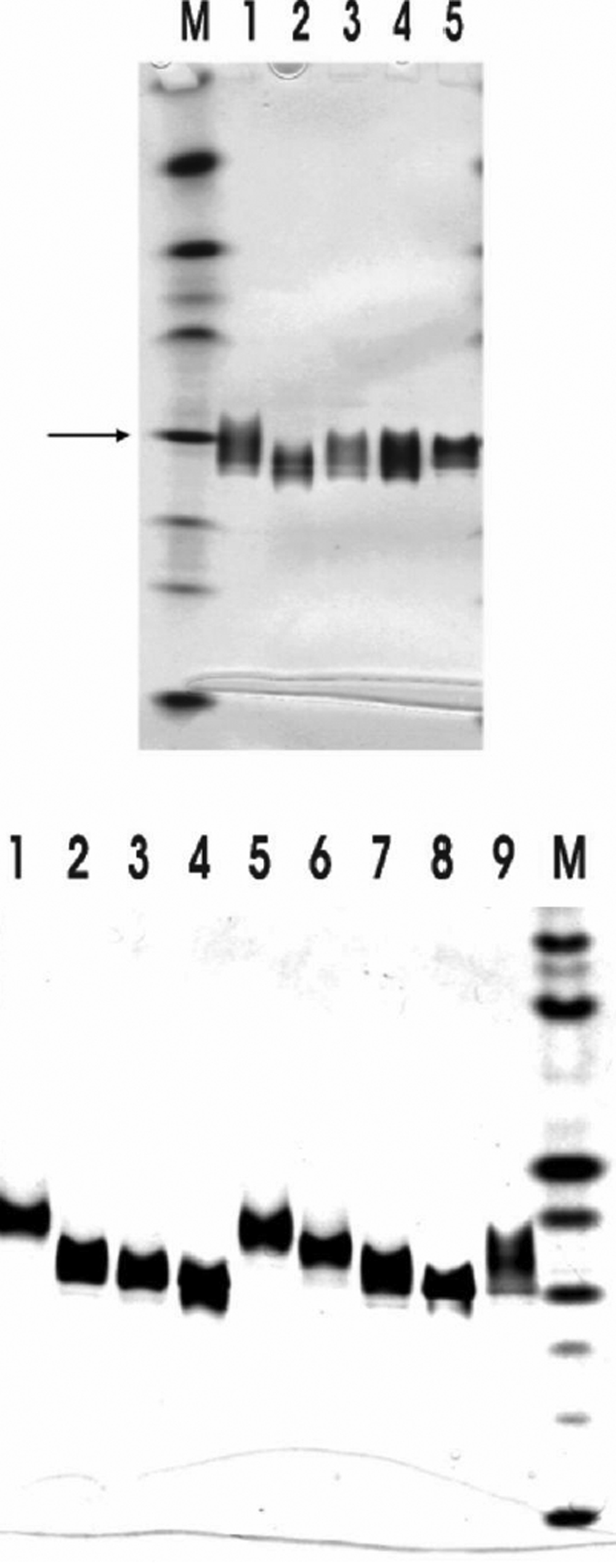
 Figure 3 of
Liao, Mol Vis 2009; 15:1429-1444.
Figure 3 of
Liao, Mol Vis 2009; 15:1429-1444.