Figure 4. M30 CytoDeath staining of retinal sections. Paraffin sections of eyes from non-diabetic rats (A), non-diabetic rats treated with etanercept (B), diabetic rats (C), and diabetic rats treated with etanercept (D) were assessed with M30 staining. Sections were subsequently counterstained with hematoxylin. Insert shows an overview of
a diabetic retina with the ganglion cell layer (above), the inner nuclear layer (middle, light blue) and the outer nuclear
layer (below, dark blue). Numbers of positive endothelial cells were counted in each of 12 sections and expressed as percent
of total endothelial cells. Treatment with etanercept reduced endothelial cell apoptosis and caspase activity as determined
by M30 staining.
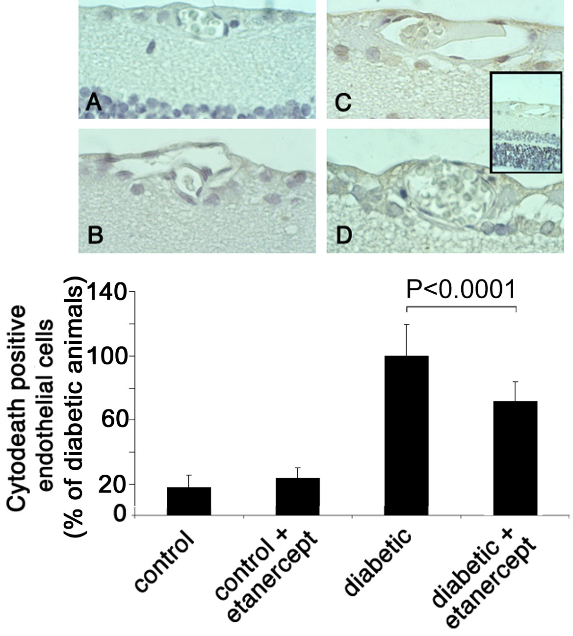
 Figure 4 of
Joussen, Mol Vis 2009; 15:1418-1428.
Figure 4 of
Joussen, Mol Vis 2009; 15:1418-1428.