Figure 2. Fundus photographs of the patient with RX mutation, R66T. Right fundus with small typical coloboma at the inferior edge of the nerve that is vertically elongated.
Note the pattern of exit of the inferior arcade vessels, indicating the presence of the coloboma.
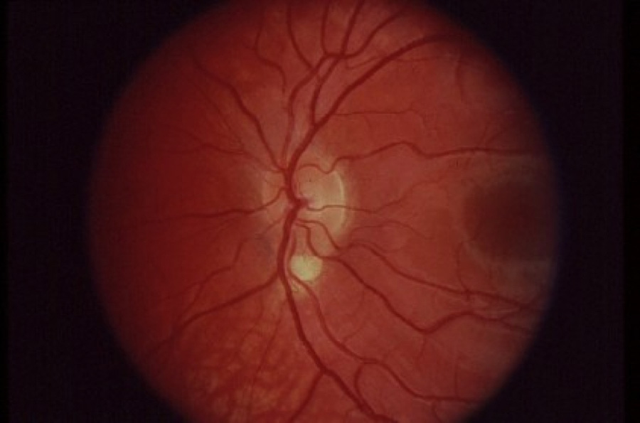
 Figure 2 of
London, Mol Vis 2009; 15:162-167.
Figure 2 of
London, Mol Vis 2009; 15:162-167.