Figure 3. Retinal microdystrophin
expression in gene transferred 3-month-old mdx3cv
mouse. Subretinal delivery of an AAV-9 human microdystrophin vector
resulted in efficient OPL transduction in mdx3cv
mouse retina. A-F are from mdx3cv
mouse eyes, and G is from a C57BL/6J mouse eye. A-C
are representative serial sections from an eye infected with AAV-9
microdystrophin vector. D-F are representative serial
sections from an eye mock-infected with HEPES buffer. A, D show
retinal structure of mdx3cv mouse (H&E
staining). B, E show immunostaining with the Dys-3 antibody,
which recognizes microdystrophin. At 5 weeks after subretinal
injection, microdystrophin expression was evident in the injected
retina (B), but not in the mock-infected eye (E). C
and F display immunostaining with the Dys-2 antibody, which
recognizes endogenous dystrophin. Neither the AAV-9-infected nor the
mock-infected eye showed endogenous dystrophin expression. G
shows immunostaining with the Dys-2 antibody on the C57BL/6J
retina. Dystrophin expression is seen in the outer plexiform layer
(OPL). Arrows point to the OPL. Abbreviations: inner nuclear layer
(INL), outer nuclear layer (ONL).
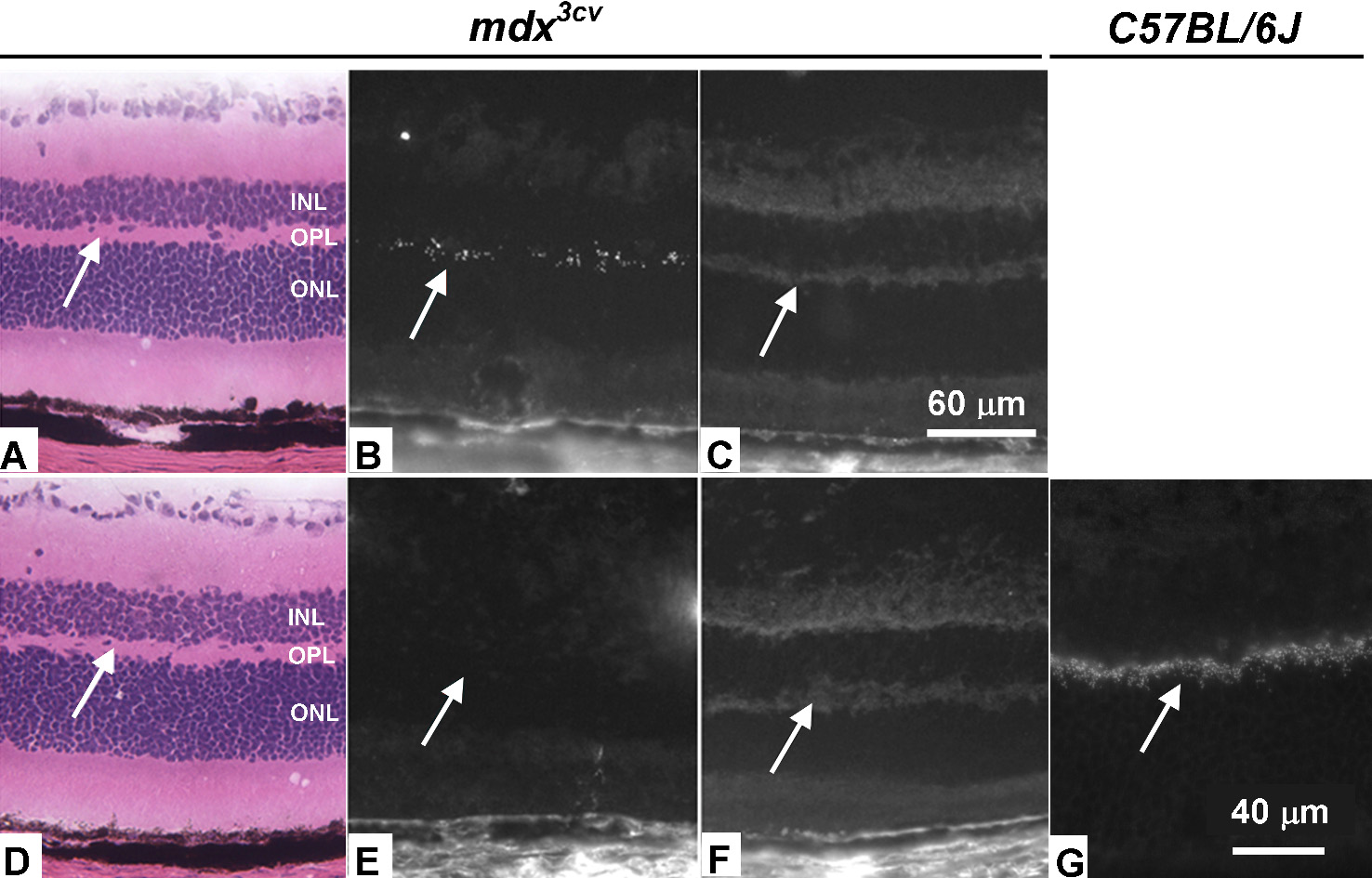
 Figure 3 of Lei, Mol Vis 2009; 15:1374-1382.