Figure 2. Retinal eGFP expression after
subretinal delivery of an AAV-9.CMV.eGFP vector in a 3-month-old C57BL/6J
mouse. AAV-9 led to widespread (from the injection site, which is close
to the posterior pole, to the peripheral retina) and throughout (from
the outer retina RPE layer to the inner retina RGC layer) eGFP
expression in the mouse retina. The eGFP expression pattern near the
injection site and in areas remote from the injection site was similar.
The pictures were taken at approximately 300 to 500 µm away from
the injection site. A-C were taken under the same
exposure conditions. D-H were taken with a shorter
exposure time. A is a section from a control eye. B
shows eGFP expression in the retina, and C is a merged picture
of B and DAPI staining. B and C show GFP
expression in the RPE, photoreceptors (including the outer and inner
segment), ONL, OPL, Müller cells in the INL, IPL and RGC layer. Because
of a shorter exposure time (D), eGFP expression was only seen in
the outer retina including RPE and the photoreceptor layer. No
expression was observed in the inner retina. E is a merged
picture of eGFP expression and CtBP2, DAPI staining. In the distal
portion of the OPL, colocalization of eGFP expression and CtBP2
staining is evident (arrow). F, G, and H are enlarged
pictures of the OPL. F. eGFP expression is evident in the
distal portion of the OPL, which is beyond the photoreceptor nuclei (in
blue). G shows CtBP2 staining (arrow) in the photoreceptor
terminals. Panel H is a merged picture of eGFP expression and
CtBP2, DAPI staining. eGFP expression overlaps with CtBP2 in the
photoreceptor terminals in the distal portion of the OPL The
calibration bar is 50 μm for A-E, and 20 μm for F-H.
Abbreviations: inner nuclear layer (INL), inner plexiform layer (IPL),
outer nuclear layer (ONL), outer plexiform layer (OPL), retinal
ganglion cell (RGC), retinal pigment epithelium (RPE).
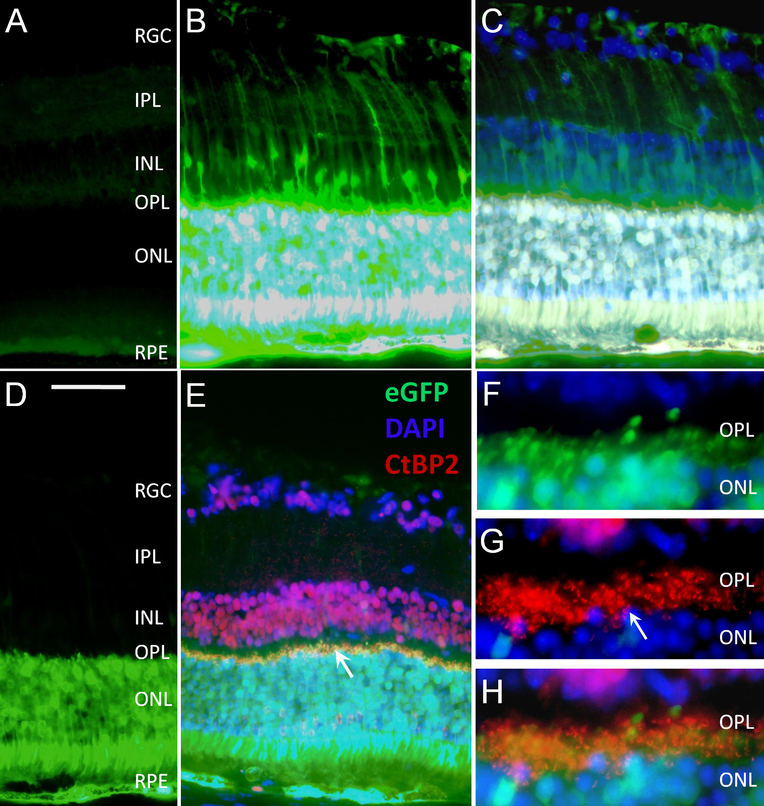
 Figure 2 of Lei, Mol Vis 2009; 15:1374-1382.