Figure 5. The predicted secondary
structures of the wild-type and the mutant αB-crystallin. The predicted
secondary structures of the wild-type form (A) and the mutant
form (B) are shown. The target sequences are labeled with white
circles. White: helix, Yellow: sheet, Pale blue: coil.
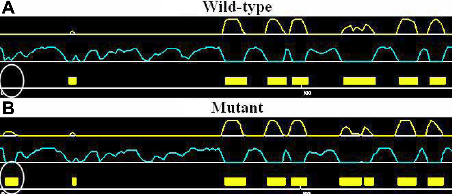
 Figure 5 of Chen, Mol Vis 2009; 15:1359-1365.