Figure 2. Confirmation of the mutation by
PCR-RFLP method. The positions of the Fnu4HI restriction sites (GC/NGC)
in the target sequence are represented (A). The schematic
overviews show that one Fnu4HI restriction site was disrupted in the
mutant form as a result of the mutation. In the wild-type form, there
are two major fragments of 221 bp and 81 bp. In the disease form, one
of the Fnu4HI restriction sites is disrupted, resulting in a longer
fragment of 110 bp (boxed region). This longer fragment can only be
observed in the affected family members (B). M, DNA Marker; Lane
1, unrelated normal control; Lane 2, senile cataract patient; Lane 3,
unaffected member of the family; Lane 4 and 5, proband and his son;
Lane 6, undigested PCR product.
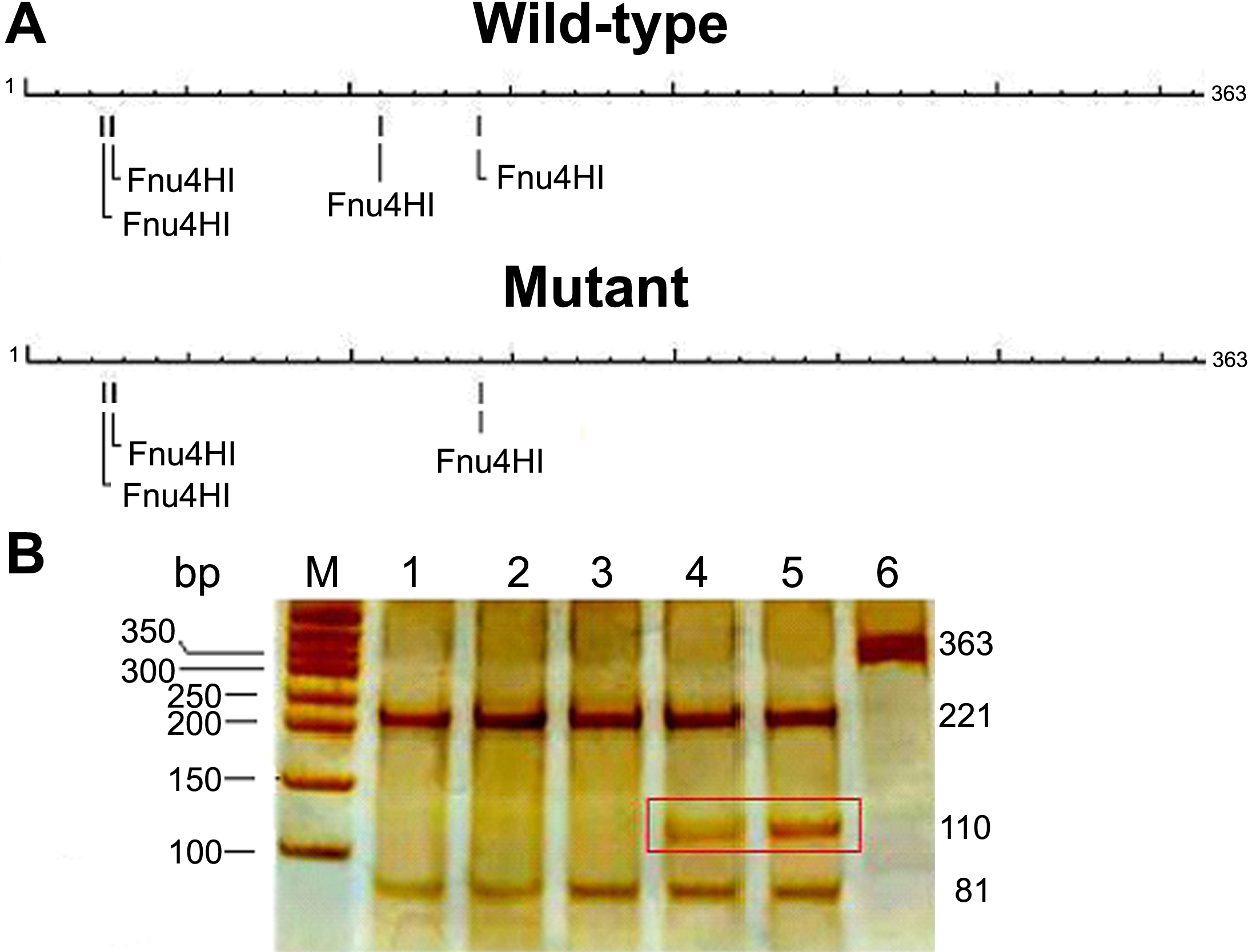
 Figure 2 of Chen, Mol Vis 2009; 15:1359-1365.