Figure 9. Confocal laser scanning
microscopy on choroidal neovascularization membranes. Images display
choroidal neovascularization (CNV) membranes of 2-month old C57BL/6
mice after subretinal injection of retinal pigment epithelium (RPE)
cells and microbeads. Tissues section with CNV lesions were stained
with antibodies against various cytokines and cytokeratin (CK) 18 (left
column) secondary Abs only; middle is with antibody to cytokine only;
right shows merged image. Cytokine expression revealed a time-dependent
release of vascular endothelial growth factor (VEGF; A-C: post
inoculation [PI] day 3), matrix metalloproteinases (MMP)-2 (D-F:
PI day 7), MMP-9 (G-I; PI day 10), and monocyte chemoattractant
protein (MCP)-1 (J-L; PI day 14) by CK 18-positive RPE cells.
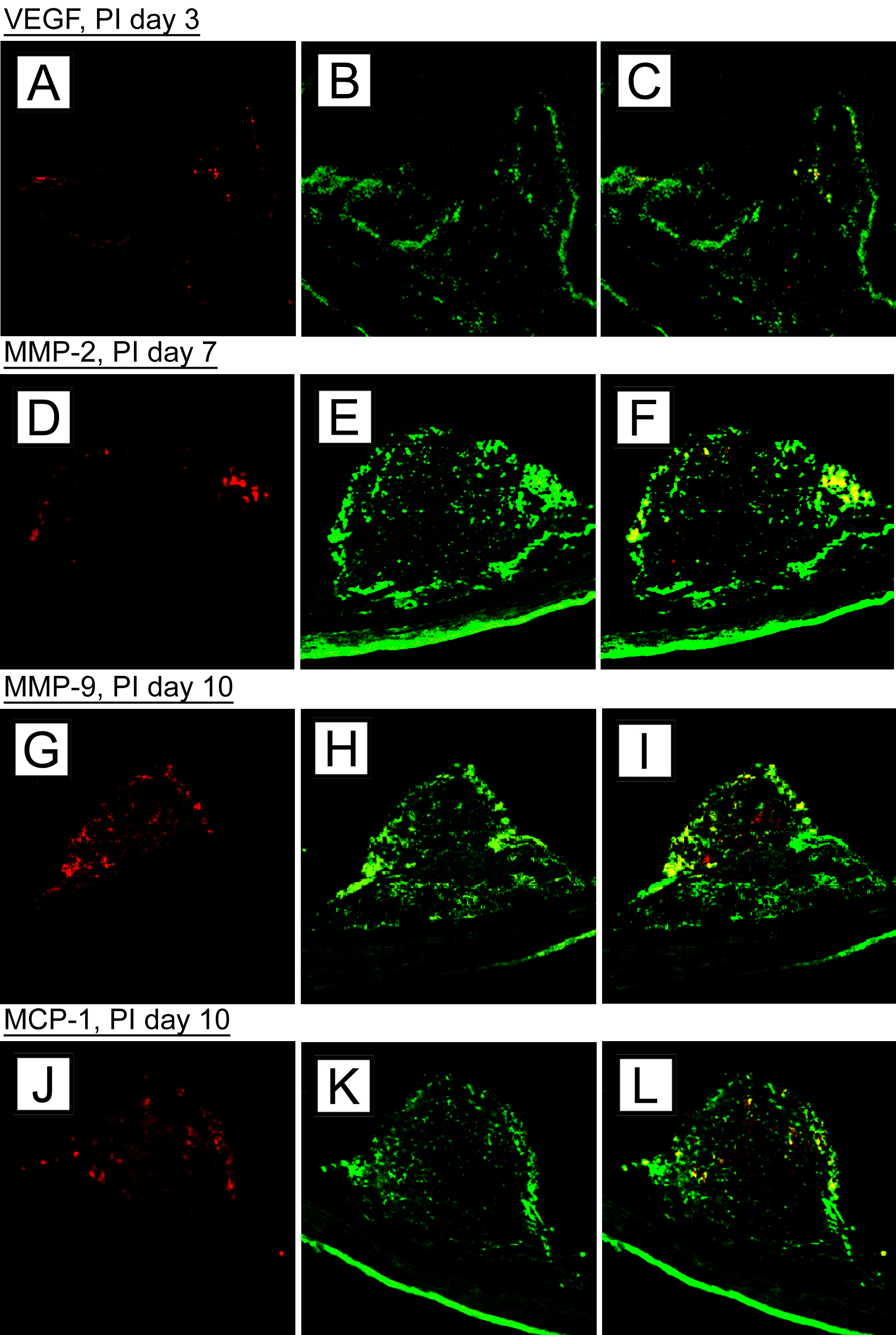
 Figure 9 of Schmack, Mol Vis 2009; 15:146-161.