Figure 7. Representative hematoxylin and eosin stained cross-sections of subretinal choroidal neovascularization lesions. Membranes
extended into the subretinal space between the retinal pigment epithelium (RPE) monolayer and the photoreceptor outer segments
of the neurosensory retina. Focal damage of Bruch’s membrane and choriocapillaris (asterisk) were present in all lesions (A, image magnification 20X). Subretinal injections were often accompanied by mild inflammation and choroidal bleeding as shown
in representative CNV membrane 3 days after subretinal injection of RPE cells (A, arrows). Microbeads and RPE cells were usually equally distributed within the CNV membranes (B, main image magnifaction 40X; PI day 7). In contrast, CNV membranes induced by subretinal injection of microbeads often demonstrated
accumulation of microbeads at the inner surface of the lesion (B, insert magnification 20X; PI day 7). Newly formed blood vessels, which stained positive for von Willenbrand factor (C, insert bottom right, asterisks, magnification 40X; PI day 10), arose from the choroid and were present in CNV lesions from
PI day 3 thru 21 (C, main image and insert bottom left, arrows, magnification 40X; PI day 3 and PI day 21 respectively). Initially, CNV surface
was free of pigmented RPE cells (D, insert bottom left, PI day 3, magnification 10X). Over time (PI day 7 to 10) spindle-shaped RPE cells (arrows) started growing
over the CNV membranes (D, insert bottom right, PI day 10, magnification 20X) and covered the lesions almost completely by PI day 21 (D, main image, PI day 21, magnification 20X).
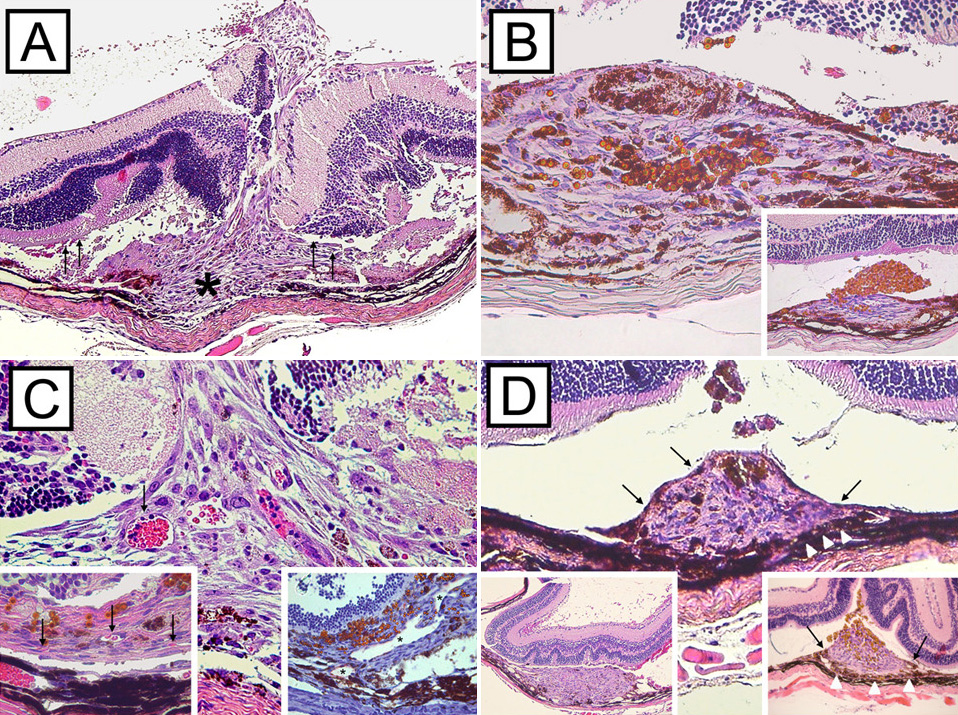
 Figure 7 of
Schmack, Mol Vis 2009; 15:146-161.
Figure 7 of
Schmack, Mol Vis 2009; 15:146-161.