Figure 6. Fluorescein angiograms of a
subretinal choroidal neovascularization membrane. Fundus photographs of
a mouse eye with a choroidal neovascularization membrane (asterisk) 14
days after subretinal injection of microbeads. The original subretinal
injection site is indicated by arrowheads (A-C). Sequential
fluorescein angiograms (B,C) demonstrate fluorescein leakage
(white arrows) around the injection site about 120 to 180 s after
intraperitoneal installation of the fluorescein dye.
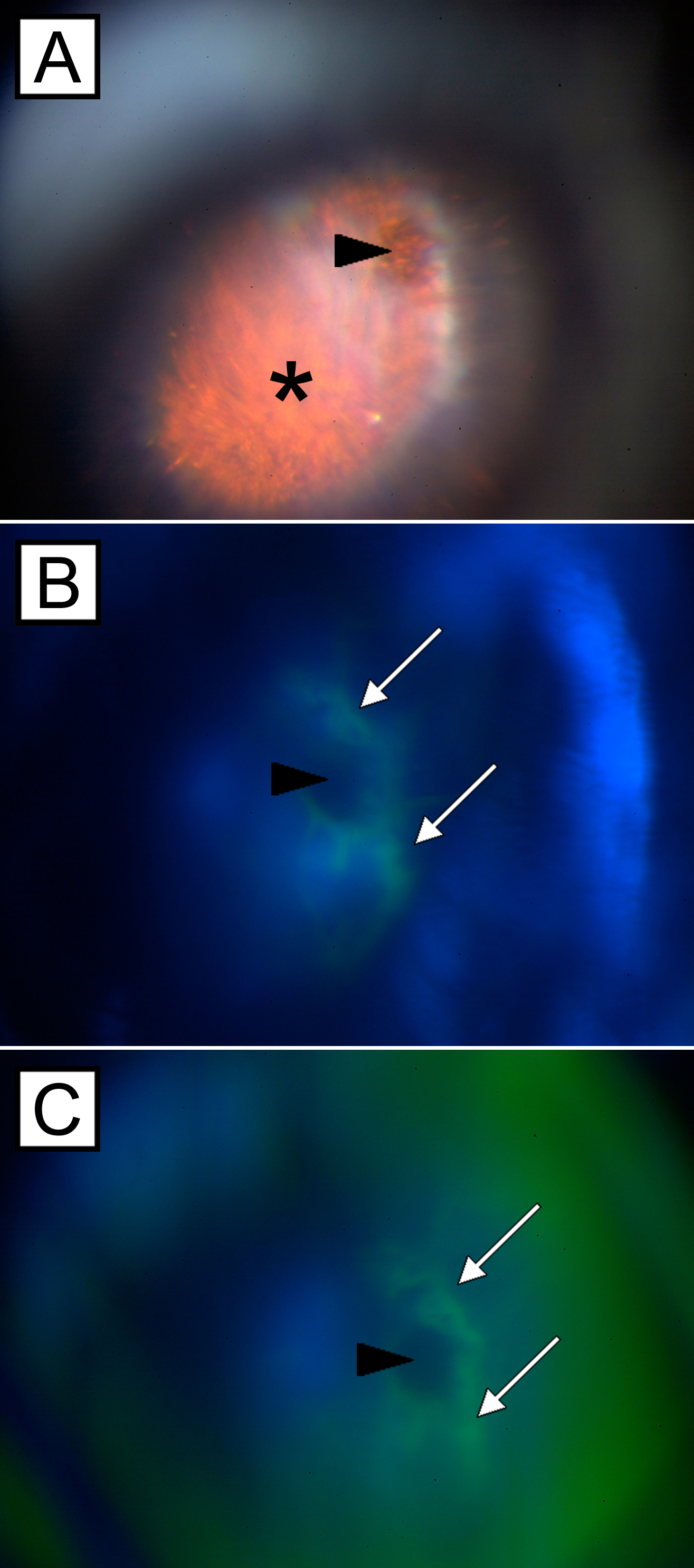
 Figure 6 of Schmack, Mol Vis 2009; 15:146-161.