Figure 4. Illustration of the choroidal neovascularization thickness measurement technique. The photomicrograph demonstrates a representative
choroidal neovascularization (CNV) membrane CNV 14 days after subretinal injection of retinal pigment epithelium (RPE) cells.
The distance between the outer surface of the choroid and the inner surface of the CNV, defined as T, and the distance between
the inner border of the choroid and the RPE monolayer next to the CNV, defined as C1 and C2, respectively, are indicated by
arrows. The relative thickness of the CNV membrane, defined as R, was calculated as follows: R=(T-C)/C. C=(C1+C2)/2. Original
magnification is 40X.
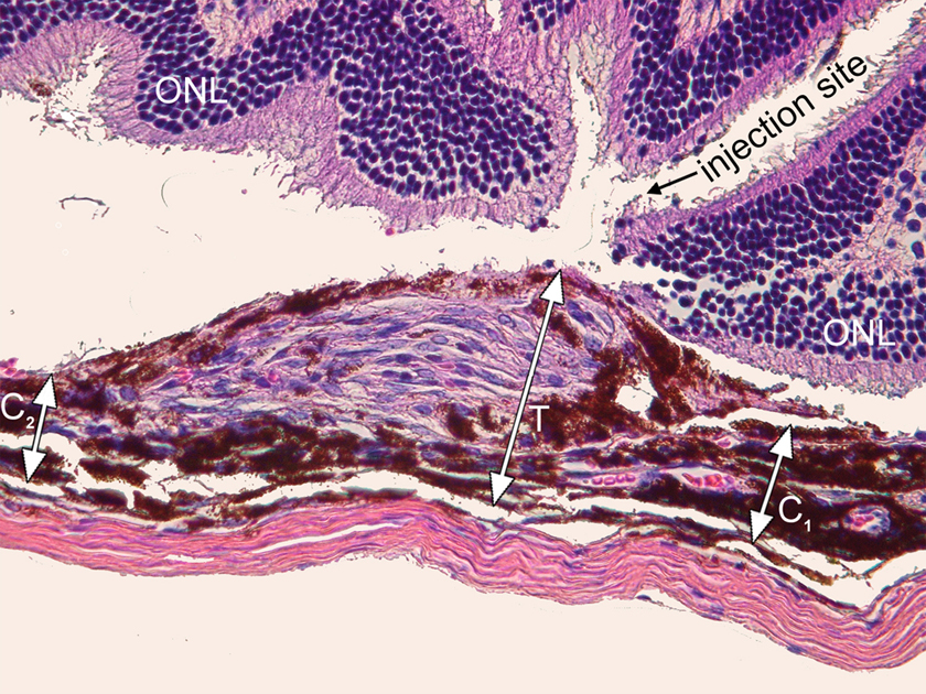
 Figure 4 of
Schmack, Mol Vis 2009; 15:146-161.
Figure 4 of
Schmack, Mol Vis 2009; 15:146-161.