Figure 3. Mouse eyes showing retinal detachment after subretinal injection. Fundus photographs demonstrate circumscribed areas of retinal
detachment (asterisks) following subretinal injection of retinal pigment epithelium (RPE) cells and microbeads (A,B). Occasionally, microbeads (B, arrowhead) and RPE cells could be observed in the vitreous cavity next to the injection site (A, arrow).
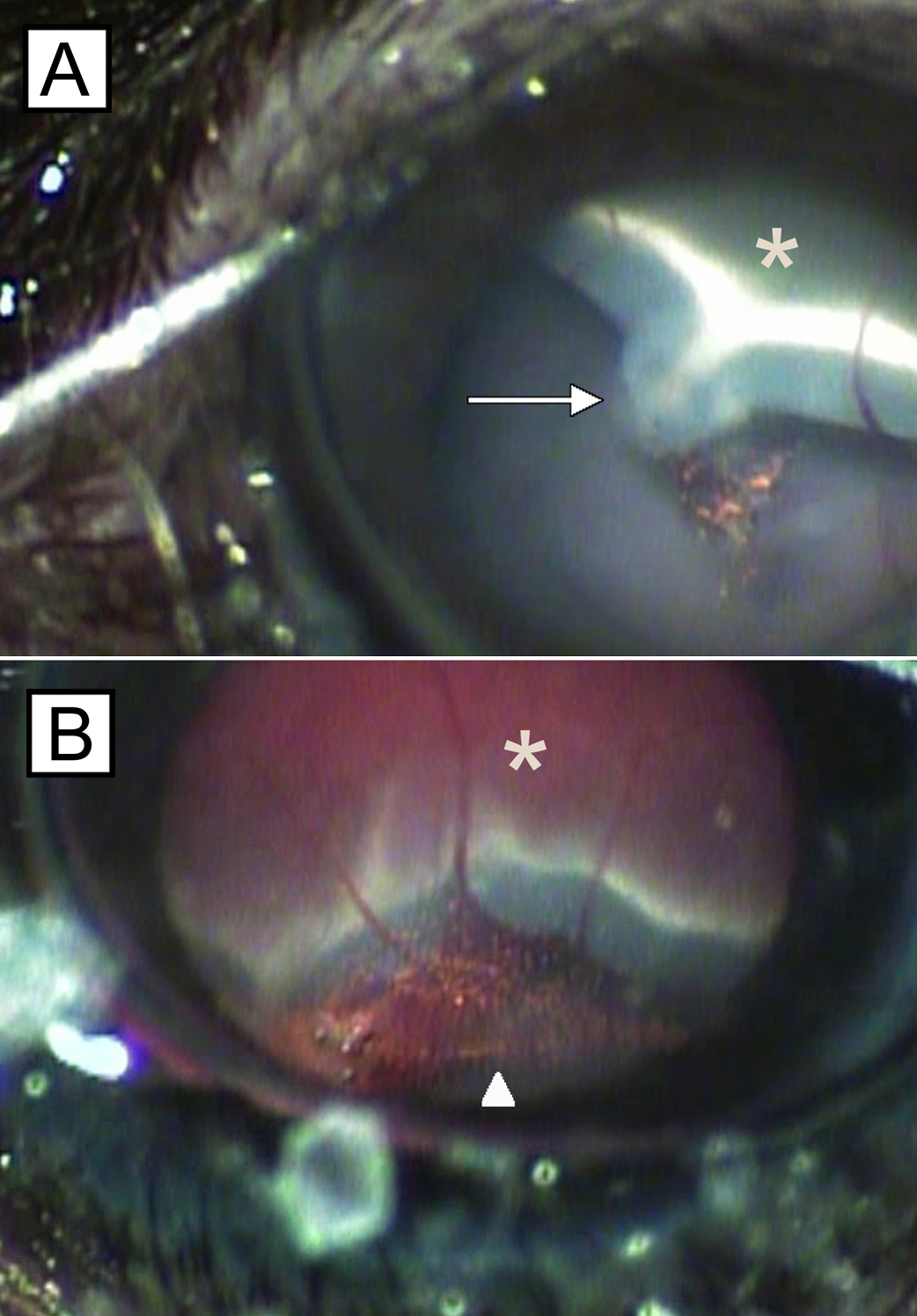
 Figure 3 of
Schmack, Mol Vis 2009; 15:146-161.
Figure 3 of
Schmack, Mol Vis 2009; 15:146-161.